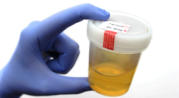
A basic but essential component of routine urinalysis includes an assessment of the physical properties of urine: colour, clarity, and USG. This is followed by a dipstick (chemical) examination (glucose, bilirubin, urobilinogen, ketones, blood, pH, and protein), and finally, a microscopic examination of the urine sediment.
The dipstick pads for leukocytes, USG, and nitrite are not considered accurate in veterinary species and are not reported.
Colour:
- Examine for colour through a clear container utilising a good light source and a white background.
- Colourless to straw coloured in cats and dogs is normal.
- Horses and cattle may have darker yellow urine due to dietary pigments, rabbits and guinea pigs may have darker yellow to reddish brown.
- Horse urine may turn red or brown with storage or exposure to snow. It is thought to be from breakdown products of catecholamines and is not associated with a pathological process.
- Red is caused by erythrocytes, haemoglobin and myoglobin.
- Orange is caused by bilirubin.
- Yellow-green or yellow-brown is caused by biliverdin and bilirubin.
- Various drugs may cause colour changes (unpredictably).
- Pigmenturia can hinder interpretation of other reactions, particularly pH, protein, and ketones.
Differentiating haematuria from haemoglobinuria from myoglobinuria
| Haematuria | Haemoglobinuria | Myoglobinuria | |
| PCV | WRI | Decreased | WRI |
| Plasma/serum colour | Colourless / straw | Pink to red | Colourless / straw |
| Urine colour (pre-spin) | Pink to red | Pink to red | Dark red / brown |
| Urine blood (haem) | + | + | + |
| RBCs in urine sediment | + | – | – |
| AST and CK | WRI | WRI | Increased |
WRI = Within reference interval
Clarity:
- Described in terms such as clear, hazy, slightly cloudy, cloudy and turbid.
- Freshly voided normal urine is usually clear to very slightly cloudy in cats and dogs.
- Horses, rabbits, guinea pigs, and goats may have mildly turbid urine (mucus +/- calcium carbonate crystals).
- Turbidity is due to suspended particles in the urine such as cells, crystals, bacteria, casts, sperm and lipid droplets. The USG is not affected but reading the line on the handheld refractometer may be more difficult.
Urine specific gravity (USG):
- Urine SG is a measurement of the density of urine compared to water. For routine clinical purposes, it is determined using a refractometer.
- Large amounts of protein and glucose will alter the urine SG and should be taken into consideration when interpreting the urine SG results. Urine SG will increase by between 0.003 and 0.005 for every 10g/L of protein and 10 g/L of glucose present.
- Where possible the USG measured before fluid administration.
- Allow refrigerated urine to warm to room temperature for 20 minutes. Cold urine falsely increases USG.
- USG helps verify that an azotaemia is due to renal failure rather than dehydration. Interpret USG along with:
- Physical examination estimation of hydration status
- Serum urea, creatinine, and albumin
- Amount of urine present in bladder
- Dilute urine (SG < 1.008) may cause osmotic lysis of cells
- There is no “normal” USG. Values that indicate adequately vs inadequately concentrated urine are reasonable guidelines rather than mathematically exact limits.
- On a randomly drawn sample, “adequate” concentrating ability is considered to be indicated by:
- Dog ≥1.030
- Cat ≥ 1.035
- Horse & cattle ≥ 1.025
- Rabbits 1.003 – 1.036, average 1.016
- Guinea pigs with uroliths mean USG was 1.007 – 1.023 in one study.
- Causes for urine that is poorly concentrated include endocrinopathies e.g. Cushing’s and diabetes mellitus, hypercalcaemia, E.coli urinary tract infections, pyometra, liver failure, drugs e.g. steroids, diuretics.
- Early renal insufficiency may result the loss of concentrating ability prior to the onset of azotemia. In this instance, checking serum SDMA concentrations may help detect early loss of renal function.
- It should be noted that SDMA concentrations can become elevated prior to the loss of concentrating ability, so careful monitoring of urine SG and renal parameters in these patients is advised.
- Cats may maintain some concentrating ability early in renal failure i.e. may be azotemic but not yet isosthenuric.
Conversion of USG results from a medical refractometer to feline urine USG values:
Conversion calculation: Feline USG = (0.846 x medical refractometer USG) + 0.154
| Medical Refractometer | Feline SG |
| 1.005 | 1.004 |
| 1.010 | 1.008 |
| 1.015 | 1.013 |
| 1.020 | 1.017 |
| 1.025 | 1.021 |
| 1.030 | 1.025 |
| 1.035 | 1.030 |
| 1.040 | 1.034 |
A Note on Refractometers
- Temperature-compensated veterinary-specific refractometers should be used, refractometers calibrated for human urine (“medical refractometers”) give erroneous results for cats, guinea pigs, and rabbits
- See conversion chart below for converting feline USG if a medical refractometer is used
- Quality control – distilled water provides an inexpensive zero calibrator; mid and high level calibrators of 5% sodium chloride (1.022 +/- 0.001) and 9% sucrose (1.034 +/- 0.001) can be used respectively.
- Temperature compensated refractometers may never need adjustment, but should be assessed periodically.
Urine Dipstick Results:
- Dipsticks consist of chemically impregnated test pads attached to a plastic strip. When the test pad is immersed in urine, a colour producing chemical reaction occurs. Results are generally semi-quantitative.
- Analyse a fresh, well-mixed urine sample, or if refrigerated, allow sample to come to room temperature.
- Submerge test strip briefly into the urine sample (no longer than 1 second) and drain excess urine by blotting the lateral edge of the dipstick against absorbent paper, ensuring urine doesn’t flow from one test pad to another.
- Compare test pad colour reactions at the specified time intervals using the dipstick analysis chart.
- Results from leucocytes, USG and nitrite are not valid in veterinary species.
Urine Glucose
Glucose is a small molecule that passes freely through the glomerulus into the ultrafiltrate, where it is reabsorbed by the proximal renal tubular cells. Glucosuria can be transient e.g. stress hyperglycaemia in cats, cattle and camelids and in dogs with acute pancreatitis. It can also be persistent e.g. due to diabetes mellitus, occasionally with Cushing’s disease (due to endogenous steroids), and acromegaly. Use of certain drugs (e.g. xylazine, glucocorticoids and progesterone) and ethylene glycol toxicity may cause glucosuria. Tubular dysfunction can result in glucosuria without hyperglycaemia e.g. acquired or congenital Fanconi syndrome. Puppies <8 weeks of age may have a mild glucosuria due to tubular immaturity.
Renal threshold of glucose in SI units, with conventional units in parenthesis:
- 10-12 mmol/L Canine (180-220 mg/dL)
- 16 mmol/L Feline (280 mg/dL)
- 8 mmol/L Equine (150 mg/dL)
- 6 mmol/L Bovine (100 mg/dL)
False positive results can occur when there are oxidising agents present. An example would be collection of urine from a table or cage floor where hydrogen peroxide or chloride bleach was present.
False negative results can occur in the presence of ascorbic acid, with marked bilirubinuria and in very concentrated samples or cold samples not brought to room temperature.
Glucose increases the urine SG by 0.004 for each 55 mmol/L or 2.5 g/dL
3+ Glucose (>0.06 mmol/L or >1 g/dL) adds 0.004 – 0.005 to USG
4+ Glucose (>0.11 mmol/L or >2 g/dL) adds 0.008 – 0.010 to USG
Urine Bilirubin
With the exception of the dog, which has a low renal threshold, bilirubin is not expected in the urine of domestic mammals. Because of the low urine threshold, dogs can have a trace to 1+ reaction, especially in well concentrated samples. Male dogs also have the ability to conjugate bilirubin in their renal tubules, which can lead to a positive result. The cat renal threshold is at least 9 times higher than in dogs, and thus any bilirubin is significant in this species.
Detection of bilirubin in urine is indicative of cholestatic hepatobiliary disease, functional cholestasis (due to extrahepatic sepsis) or haemolysis (free haemoglobin is conjugated to bilirubin in the renal tubular cells). In dogs, due to the low renal threshold, bilirubinuria can precede the onset of hyperbilirubinaemia.
Occasionally, bilirubin crystals will be present in the urine sediment but the dipstick pad is negative for bilirubin. The cause of this is unknown, but the presence of crystals is likely to indicate bilirubinuria.
False positive reactions can occur in deeply pigmented urine, with the presence of metabolites of the NSAID drug etodolac and with indican, an intestinal bacterial metabolite. False negative results may occur with sample aging, exposure to UV light, nitrites, ascorbic acid.
Urobilinogen
Not routinely used in veterinary species due to unreliable results but a positive result indicates an unobstructed biliary system and that urine is fresh.
Urine Ketones
Ketones are intermediate products of fat metabolism and form secondary to excess lipid mobilisation. Their presence indicates a state of negative energy balance. The ketone bodies, acetoacetate and acetone, are detected by the dipstick ketone pad, whereas beta-hydroxyacetate (BOHB) is not. The colour change on the pad is subtle, leading to false positive trace or small reactions, especially in well concentrated urine or urine containing blood or haemoglobin.
Some specific examples of ketonuria include:
- Diabetic ketoacidosis in cats and dogs.
- Bovine ketosis and pregnancy toxaemia in ewes and camelids. BOHB is predominant “ketone” formed in these species, dipstick does not detect BOHB.
- Starvation or malnutrition, especially in immature animals, and with low carbohydrate diets.
Urine Blood
A positive dipstick reaction for blood can result from haematuria, haemoglobinuria or myoglobinuria and the presence of any of these substances in urine is abnormal.
- Haematuria can be macroscopic or microscopic and is due to the presence of intact RBC. A minimum of 5-20/uL is needed to produce a positive reaction.
- Haematuria can result from haemorrhage anywhere along the urinary tract. Some causes include infection, inflammation, trauma, rodenticide toxicity, uroliths, neoplasia and idiopathic renal haematuria. False positive results could potentially occur with contamination from the genital tract e.g. a free catch sample from a bitch in heat.
- Cystocentesis and catheterisation can induce microscopic haematuria and a trace to 1+ blood reaction on dipstick.
- If reaction is caused by haematuria, the urine sample will go from pink to yellow and a button of RBCs will form in the bottom of the tube after centrifugation of the urine sample.
- A positive blood reaction but a lack of intact RBCs in the sediment can occur with haemoglobinuria, myoglobinuria, very small numbers of RBCs, or lysis of red blood cells (common with dilute urine).
- Haemoglobinuria is caused by intravascular haemolysis and myoglobinuria by severe muscle injury due to rhabdomyolysis.
- False positive reactions can occur with bleach (home urine containers should be clean but not “sterilised” by using bleach for this reason).
Urine pH
- Many renal and extra-renal factors affect urine pH. Carnivores usually have acidic urine, whereas herbivores usually have alkaline urine, unless on a milk diet.
- Knowledge of the urine pH is important when interpreting urine sediment as alkaline urine is more likely to result in disintegration of erythrocytes, leucocytes and casts. Alkaline urine can also cause a falsely elevated pH reading on the dipstick pad.
- A false decrease in the pH pad reading can occur if urine leaks across from the protein pad.
- Normal urine pH:
- Dog and cat: 6.5 – 7.5
- Equine and bovine: 7.5 – 8.5
Some factors that influence urine pH include:
- Diet – elevated pH in dogs and cats after eating, the postprandial alkaline tide, is due to increased secretion of HCl into the stomach. Dogs that have a large proportion of their diet as vegetables, will have a high urine pH.
- Abnormal acid/base balance or tubular dysfunction.
- Age of specimen – loss of CO2 to the air occurs, raising pH.
- Presence of bacteria – urease-positive bacteria such as Streptococcus, Ureaplasma or Proteus spp. convert urea to ammonia, which will increase the pH. A persistently alkaline urine should raise suspicions for a UTI. Infection with these bacteria can lead to concurrent urolith formation as crystals precipitate out more readily in alkaline urine.
- Acidic urine in cows raises concern for displaced abomasum or upper GI foreign body.
Urine Protein
- Proteinuria may be:
- Pre-renal – also called overload proteinuria and occurs when the amount of filtered protein is in excess of the ability of the renal tubules absorptive capacity. Examples include colostral proteinuria in neonates <40 hours old; excess free light chains associated with plasma cell malignancies; haemoglobinuria and myoglobinuria.
- Renal
– renal proteinuria is defined as persistently elevated
UPCR >0.5, where pre- and post-renal proteinuria have been ruled out. It may
be of glomerular, tubular or interstitial origin.
- Glomerular proteinuria can be functional e.g. due to stress, fever, excitement and congestive heart failure OR pathological due to amyloidosis and glomerulonephritis. The proteinuria is generally moderate to marked and can result in hypoalbuminaemia and other complications if loss is prolonged or severe.
- Tubular proteinuria can either be due to reduced tubular function resulting in decreased absorption or increased excretion of proteins by damaged tubules. There are many causes including renal ischaemia due to hypoxia or hypotension, toxic insults, infectious diseases.
- Interstitial proteinuria less common and due to inflammation or haemorrhage within the kidney.
- Post-renal – lower urinary or genital tract inflammation or haemorrhage. Note that microscopic haematuria associated with cystocentesis does not usually result in haematuria.
- The protein pad on the dipstick is more sensitive to albumin than globulins and is insensitive to Bence-Jones proteins.
- Interpret results in light of the urine specific gravity and pH.
- Normal urine contains little to no detectable protein so proteinuria in the absence of inflammatory sediment or blood, i.e. a quiet sediment, is evidence of renal protein loss. Trace to 1+ results can occur in well concentrated samples of >1.030, however in dilute samples would be abnormal, as would a 2+ or more in a well concentrated sample.
- Albumin in urine increases USG by 0.003 for each 10 g/L.
- Confirm dipstick results with a urine protein:creatinine ratio as the dipstick result is only subjective. (UPCR; see Quantitative Urinalysis section below).
False positives – highly alkaline urine, highly pigmented samples, prolonged immersion of urine sample, run over between pH and protein test pad, and presence of chlorhexidine skin cleanser.
False negatives – Bence-Jones proteins are not detected due to insensitivity of the protein dipstick pad; microalbuminuria where protein concentrations are very low; highly acidic urine.

