
Urine can be collected by a variety of methods including cystocentesis, catheterisation, manual expression, voided, or from the floor, table or litter tray when necessary. State the method of collection on the history form, to aid interpretation.
Cystocentesis:
This is the collection method of choice, as it is the most sterile and therefore suitable for culture. It is generally considered to be safe, however there are some risks. The most common is microscopic haematuria, which can be hard to differentiate from pathological haemorrhage. Generally, with iatrogenic haemorrhage, there is a 1+ blood on the dipstick, urine is usually not discoloured, nor is there an increase in protein concentration. Other complications include rupture of an overly distended bladder, bladder laceration, inadvertent aspiration of gut contents and peritonitis secondary to leakage of septic urine. If urine is not obtained on the first attempt, use a new needle. Ballottement prior to cystocentesis helps to mix bladder contents, and may increase findings in sediments.
Reasons for cystocentesis:
- Urine culture – helps localise infection to bladder +/- kidney v. urogenital tract
- Helps localise hematuria, pyuria, and bacteriuria
- Avoids contamination from the lower urogenital tract and skin
- Avoids iatrogenic urinary tract infection associated with catheterization
- Part of therapeutic management of blocked cats (early stages)
When not to sample by cystocentesis:
- Insufficient volume of urine in the urinary bladder
- Overlying pyoderma, coagulopathy, local neoplasia (risk of seeding)
- Patient resists abdominal palpation and restraint
Equipment needed:
- 22-25 g needle, 1½” to 2″ (depending on patient size)
- 3-12 cc syringe for diagnostic cystocentesis
- Use clean needle for transferring to the lemon top (or red top) tube
Many techniques are used and reputable resources can be found on the internet. Ultrasound guidance is ideal, but not always possible. Dorsal recumbency and aiming for where isopropyl alcohol pools on the abdomen, is another. In cats, lateral or dorsal recumbency is preferred. In dogs, the technique can be done with the animal standing. In general, immobilise bladder against body wall. Insert needle through ventral abdominal wall and into bladder at an oblique angle, aiming to enter bladder lumen a short distance cranial to urethral junction. This location allows the needle to remain in the bladder lumen as it reduces in size, which may not happen if the needle enters close to the bladder apex.
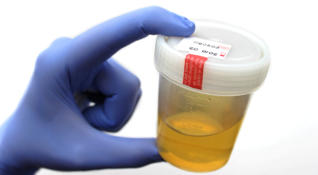
Free catch / voided urine:
This is the least invasive method of collection. Ideally, a mid-stream sample should be collected as this is less likely to contain contaminants from the vulva, prepuce or urethra e.g. cells, bacteria, hair and debris.
- Acceptable for urinalysis and sediment exam.
- Suitable for culture if no other method is possible, however state method of collection on submission form.
- Long handled ladles, foil pie plates, large cups can all be used to collect urine.
- Clean and empty litter trays can be used for cats, although Styrofoam packing peanuts are useful if a cat doesn’t like using a litter-free litter tray.
- Manual expression of the bladder is advised on patients who are sedated or anaesthetised as awake patients require greater pressure to induce micturition, often high enough to cause reflux of urine into the ureters.
Catheterisation
This method of collection requires greater technical skill, often requiring sedation or anaesthesia. Sterile technique should be followed for the patient’s urinary tract health and integrity of bacterial culture
- Blood (1+ blood on dipstick) and increased numbers of squamous epithelial cells may be seen.
- Can track materials into bladder and cause bladder infection.
- Patients that have indwelling catheters are at greater risk of bacterial UTI’s and culture is advised following catheter removal.