
A 5-month-old English springer spaniel presented with pyrexia of unknown origin, in-clinic testing showing an increased WBC count and neutrophilia. He was treated with Clavulox and his CBC had returned to normal by the last day of antibiotic therapy.
He presented again a few days later with lethargy, but a normal temperature and CBC. He responded well to corticosteroids and was placed on an immunosuppressive dose of prednisone. After another few days the owner reported PU/PD and aggressive behaviour. He was readmitted later that day in a lethargic, collapsed state, and started displaying increasingly advanced neurological signs, with several episodes over the next 24 hours, and progressively worsening seizures. The dog was euthanased on humanitarian grounds.
The body was submitted to the laboratory and a necropsy was performed. Post-mortem findings included haemorrhagic subcutaneous tissues surrounding the left eye, with dark red material within the eye (hyphaemia). Within the centre of the brain there was a soft tan area surrounding a piece of plant material – a grass awn (Figure 1).
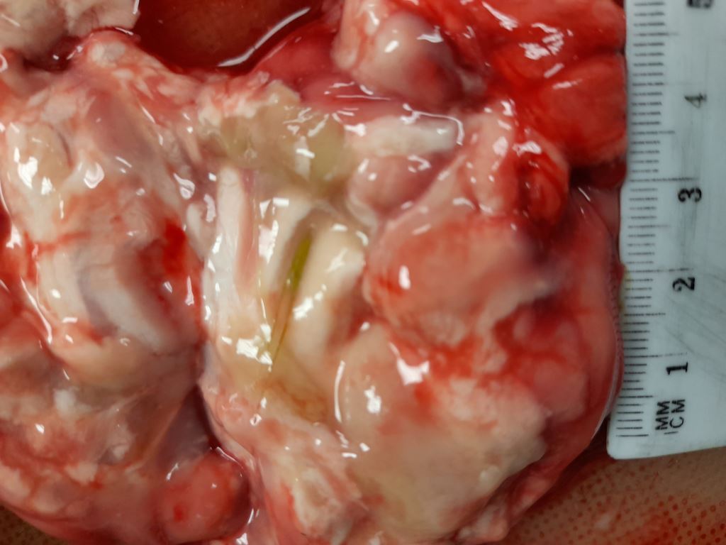
The subcutaneous haemorrhage and hyphaemia were likely secondary to trauma from the seizures. The plant foreign material (grass awn) with presumptive encephalitis explains the clinical signs. This finding is very uncommon, and the grass awn likely entered through the nasal passages.