
Species: All.
Specimen: Fixed tissue (1:10 tissue:formalin).
Container: Plain.
Collection protocol:
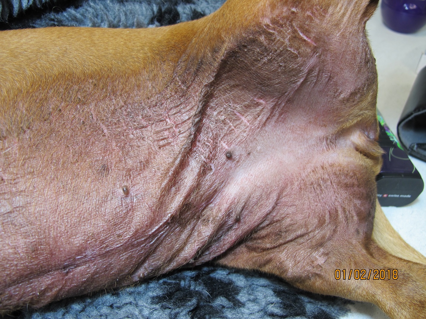
Skin biopsies are often an effective diagnostic tool. The results should be able to give you an idea of what the disease process is (e.g. inflammatory vs. neoplastic), and sometimes what agent is causing it. Even if a specific cause is not identified, by knowing if the lesion is follicular or deep dermal, neutrophil-rich or granulomatous, you can effectively reduce your differential list and focus on the most likely aetiologies. Some diseases can only be diagnosed by skin biopsy, however it is important to also recognise that biopsies are not always indicated especially for primary allergic skin disease.
- Biopsies often show non-specific changes in syndromes of allergic skin disease. Carefully consider the history, clinical signs and results of other tests before proceeding to biopsy.
- Select primary lesions to biopsy (e.g. papules, pustules, vesicles, nodules) before secondary lesions (e.g. lichenification, excoriation).
- If you are sampling an animal with inflammatory skin disease, try to sample as early in the course of the disease as possible, before the development of chronic, secondary non-specific changes. Try to sample entire fresh lesions (if small, e.g. vesicles) or the edge of larger fresh lesions.
- Conversely, if the animal has a non-inflammatory skin disease (e.g. suspected endocrine alopecia, a mass), avoid sampling the edge of the lesion or fresh lesions, and instead select the most well-developed area, since this is most likely to have classical histological changes. It should be noted it is uncommon for a skin biopsy to have changes specific to a particular endocrine disorder.
- It is important to note on the accession form any treatment the animal has received in recent weeks. Steroid drugs should optimally be withheld for 2-3 weeks prior to the biopsy. (Depot steroids might need a 6-8 week withdrawal). Antibiotics before biopsy may be helpful unless a primary infection is suspected, and culture of the biopsy required. These reduce changes due to secondary infection, important since infections can obliterate the primary lesion and sometimes mimic immune-mediated disease (e.g. pemphigus foliaceus-like dermatophytosis).
- For skin disease investigations, multiple biopsies are always advisable.
- Generally, a 6 to 8mm biopsy punch provides an adequate specimen. Smaller biopsies may be taken from the nose, pinnae and feet. Make sure the instrument is sharp to avoid distortion of the tissue, and take the punch in a continuous rotating motion rather than a “back and forth” twist.
- Ensure if possible that the lesion is in the centre of the punch biopsy, since when processed these are generally sectioned in half by default. An eccentric lesion may therefore be missed if it is not obvious at the time of processing.
- If the lesion is suspected to be deeper in the subcutaneous tissue, a punch biopsy may not penetrate deep enough. Excisional biopsy with a scalpel is indicated in this situation, or where the shearing action of the punch may damage lesions, or where there are larger vesicular and pustular lesions. These biopsies should be elliptical and a minimum of 5 x 15 mm.
- Incisional biopsies (i.e. sampling part of a mass) are often useful in cases of suspected neoplasia, where the lesion may need to be graded or staged before curative surgery is undertaken.
- In horses, full-thickness biopsies of the coronary band result in permanent hoof wall defects and therefore superficial “shave” biopsies are preferred from that area.
- It is usually not necessary to biopsy normal skin for a “control”, unless the species of animal is unusual or exotic.
- Biopsies can usually be taken with sedation, systemic pain relief and local anaesthesia of the site. If a deep dermal or subcutaneous disease is suspected it is best to avoid local injection, and to use general anaesthesia or ring blocks to prevent damage to the area. It is also important to avoid local anaesthesia if the biopsy will be cultured (lidocaine can inhibit bacterial growth).
- It is best to do as little as possible to clean the biopsy site since disinfectants, scrubbing and clipping will damage the skin and potentially remove the epidermis and valuable information (e.g. acantholytic cells in crusts, vesicles). Gently dabbing the skin with alcohol, and scissor cutting of long hair in the area is the most interference there should be.
- Care is needed in handling the biopsy specimen. It is easy to crush and distort the sample with forceps. The specimen should be gently blotted to remove excess blood. It may then be placed into a tissue cassette if very small (this reduces trauma to the sample floating in formalin during transit, and also reduces handling at the laboratory before processing).
- Punch biopsies do not curl and can be placed directly in formalin. Biopsies taken with a scalpel often need to be attached to cardboard to prevent curling. Adherence of the deep margin should occur within 60 seconds, then place the cardboard and attached sample into 10% buffered formalin. (Do not wait longer than 1-2 minutes, in order to prevent autolysis).
- Unless identification of individual sites is important all the biopsies can be placed in the same container.
General information about when this test is indicated:
Any potential neoplasm, persistent ulceration, suspected disease that can only be diagnosed by biopsy (e.g. sebaceous adenitis), any unusual dermatosis, vesicular dermatitis, any suspected disease with an expensive/difficult/dangerous treatment, any dermatosis not responding to apparently rational therapy.