
Species: All mammals
Specimen: Hairs, skin exudate on microscope slide, skin scrapings or nails.
Container: Microscope slide holder, envelope or plain container.
Collection protocol:
Wood’s lamp is an ultraviolet light instrument, often with an incorporated magnifying glass. When exposed to the UV light, hairs invaded by Microsporum canis or Microsporum equinum can give a yellow-green fluorescence (30-80% of isolates). This is due to tryptophan metabolites produced by the organisms. Fluorescence is not present in the scale or crusts, or in cultures of M. canis. There are other, less common dermatophytes that may also fluoresce. Medication can interfere with the test, for example, iodine washes can destroy the fluorescence and some bacteria and chemicals can give false positive results. Lack of fluorescence is inconclusive, and it should also be noted that most dermatophytes of horses do not fluoresce.
Hair is the specimen most commonly collected for the isolation of fungi. Using forceps, select hairs that fluoresce under the Wood’s lamp.
Alternatively the toothbrush method is effective in selecting specimens.
- Use a new toothbrush and gently comb the hair onto paper.
- Submit the brushings and toothbrush in an envelope or non-air tight sealed container for fungal isolation.
- Choice of container is important to prevent moisture build up, which allows bacterial overgrowth.
- The toothbrush comb technique is recommended in cats that are suspected carriers of M. canis.
Besides fungal isolation, which can take some time, KOH digest of hair is a rapid diagnostic screening technique for dermatophytes, which can aid in light microscope diagnosis. If negative, dermatophytosis still cannot be ruled out.
Skin samples can be submitted for fungal isolation.
- Clean the skin gently of extraneous debris with gauze soaked in 70% alcohol.
- Scrape from the periphery of the lesion (ideally a new one) and adjacent skin.
- Do not use a paraffin-coated scalpel to collect samples for microbiology.
Samples of nail and paw pad can also be cultured for dermatophytes. Since these are often heavily contaminated with micro-organisms, they should be cleaned with 70% alcohol before sampling. Samples should be selected from the concave side of the claw, or from within the claw.
Skin impression smears can be quite useful in the diagnosis of yeast infections. Simply press the slide directly onto the lesion and allow to air dry. Sticky tape preparations are difficult to work with and not recommended.
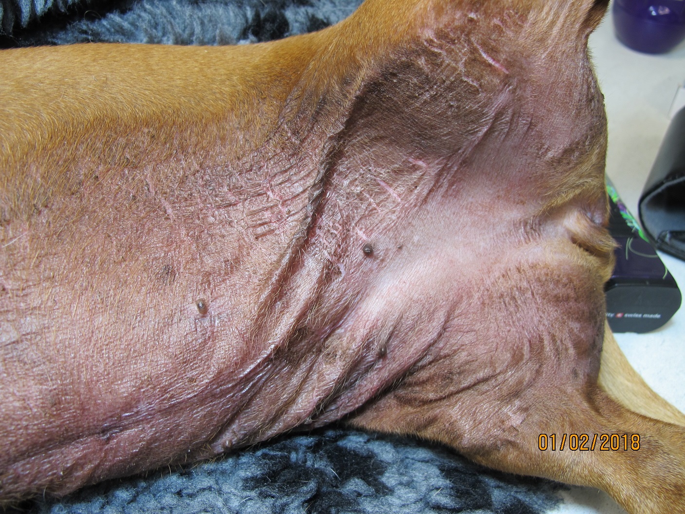
General information about when this test is indicated: Dermatoses with prominent alopecia, follicular lesions, crusting or scaling.