
Species: All
Specimen: Pustules or Vesicle contents, or scale on microscope slide or swab; fresh (unfixed) skin biopsy.
Container: Microscope slide holder, swab, E-Swab container plain container .
Collection protocol:
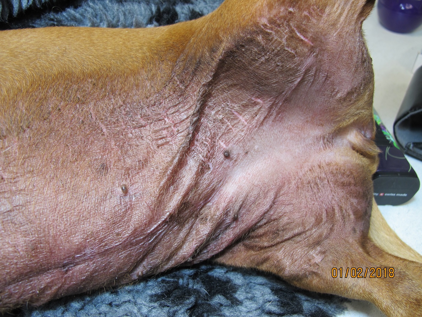
The normal skin is remarkably resistant to bacterial infection and even colonisation. When damaged, however, the environment becomes much more conducive to bacterial growth.
- Pustules, papules and vesicles can contain acantholytic cells, bacteria and the predominant granulocyte population. The presence of intracellular bacteria confirms their role in inflammation.
- Unruptured pustules/papules/vesicles need to be used. These lesions should not be prepared or sterilised before sampling.
- The sample is collected via needle puncture and/or aspiration with transfer to a sterile swab for culture, then smearing for cytology.
- Secondary bacterial infection is common in eroded, ulcerated or crusted types of lesions, and culturing these is seldom helpful.
- Culture of fresh skin biopsies may be indicated for plaques, nodules, fistulous tracts, deep lesions or cellulitis. (These sites should be sterilised before surgery and the superficial epidermis excised prior to submission ). It is helpful to also aspirate these for cytology at the same time.
- Sticky tape preparations of surface material are difficult to work with and NOT recommended.
The majority of primary bacterial skin infections are caused by coagulase positive Staphylococcus. Occasionally Proteus sp., Escherichia coli or Pseudomonas sp. are involved, usually as secondary invaders. In cats, bacterial infections are less common and usually involve Rhodococcus sp., Pasteurella sp. or ß-haemolytic Streptococcus.
Rare infections are caused by anaerobes or higher bacteria including Nocardia, Actinomyces and mycobacteria (e.g. feline leprosy). Unstained direct smears and fresh skin biopsies can be submitted for cytology and culture if one of these infections is suspected. Mycobacteria in particular can be very slow-growing and therefore these infections are usually initially diagnosed by cytology or histopathology. Follow-up testing to identify the species of mycobacteria is available in Melbourne by PCR.
General information about when this test is indicated: Dermatoses with prominent pustules, vesicles, exudate, cellulitis, nodules or fistulous tracts.