
CRISTINA GANS
Getting a diagnosis in an abortion outbreak can be challenging. Less than 50% of abortions investigated result in a diagnosis. A variety of tests are available at Gribbles Veterinary (including PCR, histology, microbiology and serology) which enhances our ability to obtain a diagnosis. However, this can also be confusing in terms of sample collection.
To necropsy or not to necropsy?
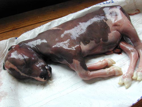
When presented with an aborted fetus, there is the option to send the fetus to us for necropsy and sampling. Please contact your local laboratory before sending fetuses. Only our Auckland, Palmerston North and Dunedin laboratories have pathologists on-site to perform post-mortems.
Which samples to take?
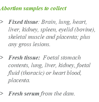
If you do perform a field necropsy, getting the recommend fixed and fresh samples can maximise your chances of obtaining a diagnosis. A list of recommended samples for bovine and ovine abortions can be found below.
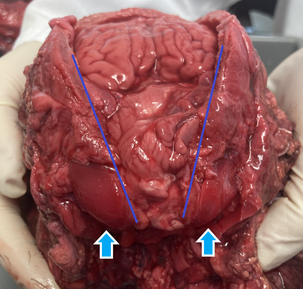
Brain may be a difficult sample to collect as it can be soft or autolysed. Ideally remove the brain intact by removing the calvarium (Figure 1). However, even if the brain is autolysed, soft or fragmented, it can still be useful diagnostically. Placing the brain into formalin will allow it to harden, even if you have to resort to “pouring” it in.
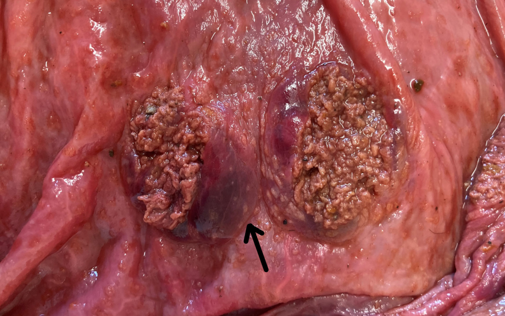
Placenta is an important sample to include if it is available. In some cases, the only gross or histological lesions are evident in the placenta. Take multiple sections of cotyledonary and non-cotyledonary placenta, as lesions of placentitis can have a patchy distribution. For example, the mycotic abortion in Figure 2 displayed areas of necrosis that affected some cotyledons or parts of cotyledons.
Skin may sometimes have plaques due to fungal abortion. It’s worth having a look at the skin and taking unusual lesions for histology. Similarly, eyelids can display histological lesions suggestive of Ureaplasma or fungal abortion in cattle.
It’s important to note that gross lesions are rarely seen during an abortion necropsy. Some lesions may be due to autolysis or may be a variation of normal (such as amniotic plaques or adventitial placentation). If unsure, its’s always best to collect a sample for histology. Photos of lesions with the submission are also helpful.
Which test to select?
The selection of tests ideally should be guided by herd/ flock history, vaccination status, any illness noted in the dam and farm environment.
Histology is a good test to start an abortion investigation. While it doesn’t often provide a specific diagnosis, it can provide information to direct other ancillary testing such as PCR or culture. Alternatively, PCR panels can provide a rapid and accurate diagnosis if a particular organism is suspected. A pathologist can help guide the selection of tests in an abortion investigation if required.
References:
Njaa, Bradley L., ed. Kirkbride’s diagnosis of abortion and neonatal loss in animals. John Wiley & Sons, 2011.
Gribbles Veterinary. Samples to collect for abortion investigations. Paws, claws & udder things, July 2019.