
With the increasing introduction of in-clinic haematology analysers, we have started to see a variety of requests for haematology services including complete blood counts (CBC), total WBC plus differentials, differentials alone and smear examination alone.
When obtaining a CBC, the simplest part of the procedure is to run the blood sample through an analyser and obtain the RBC parameters and a total WBC count, with or without a machine generated differential. This is, of course, based on the assumption that the analyser is working correctly and is giving accurate results. Most analyser technology was initially developed for use in humans and extrapolated to veterinary species. Therefore, the technology is not always adept at making important differentiation of cell types, especially when abnormalities are present. It is imperative to manually review most blood films in veterinary medicine, especially in ill animals.
Information not provided by analysers:
Accurate differentials
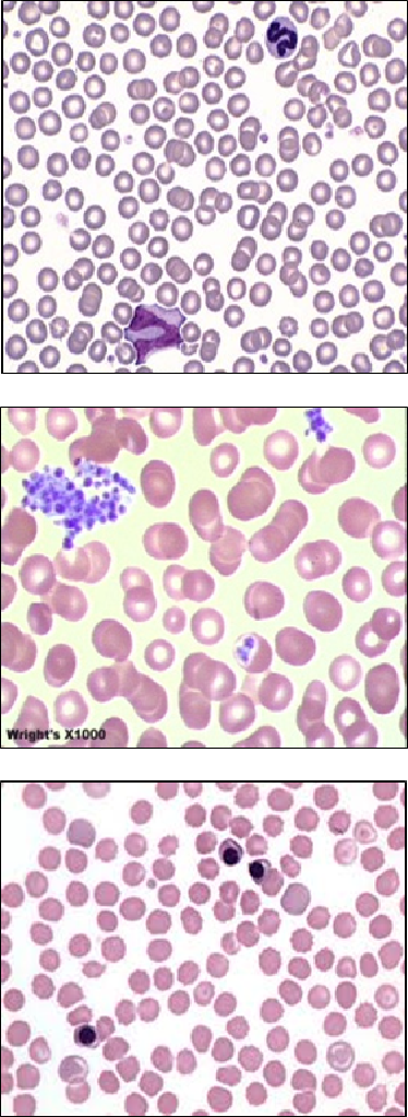
All haematology analysers can produce inaccurate white cell differentials, especially when significant abnormalities are present. Subtle but significant abnormalities in cell classification may occur, even though machine cell counts and differentials ultimately fall within the reference interval. Additionally some analysers only provide a total granulocyte count such that neutrophils, eosinophils and basophils and probably also mast cells are all counted as granulocytes. This means an animal may have an eosinophilia, basophilia or mastocytosis that will not be detected unless a differential is carried out using light microscopy. Blast cells will also not be identified or counted separately by haematology analysers.
Left shift and indicators of inflammation
The presence of Howell Jolly bodies, Heinz bodies, basophilic stippling, RBC parasites such as Mycoplasmas and various abnormal RBC shapes are not noted. These changes must be identified microscopically and may provide critical clues as to the nature of the anaemia for example spherocytes and agglutination in immune-mediated haemolytic anaemia.
RBC morphology
The presence of Howell Jolly bodies, Heinz bodies, basophilic stippling, RBC parasites such as Mycoplasmas and various abnormal RBC shapes are not noted. These changes must be identified microscopically and may provide critical clues as to the nature of the anaemia for example spherocytes and agglutination in immune-mediated haemolytic anaemia.
Platelet numbers
Large platelets may be counted as RBCs which can lower the automated platelet count. Platelet clumping routinely results in erroneously low platelet counts and has been responsible for misdiagnosing immune mediated thrombocytopenia. Microcytic RBCs may also be misclassified as platelets causing a true thrombocytopenia to be missed in rare instances.
Nucleated red blood cells
These cells are typically counted as lymphocytes by in-clinic analysers, which can result in erroneous lymphocytosis.
Additional points to consider:
Blood smear examination
The quality of a blood smear is vital to correct interpretation. Poorly made films can lead to incorrect differential counts and poor assessment of erythrocyte morphology, including misdiagnosis of spherocytes and immune mediated haemolytic anaemia. If practitioners choose to send in blood smears alone for evaluation, the quality of your interpretation will be dependent upon your ability to produce a good quality blood smear. Assessment of morphology of freshly made blood films is still vital to appropriate interpretation of some erythrocyte and leukocyte morphological changes, so as well as a freshly made blood film, an EDTA blood sample is also recommended (so additional blood smears can be prepared if necessary).
Total WBC count
A total WBC count is required to accurately produce absolute numbers of the various leukocyte types present. The percentage of a particular cell type may seem high or low on a differential count, but interpretation of true elevations and decreases in white blood cell numbers relies on the absolute count.
Differential and cell morphology
The most skilled and time consuming part of the CBC is the differential and smear examination for RBC and leukocyte morphology. This assessment relies on the ability to recognise subtle differences in cells and to correctly identify cells rarely seen in circulation. As these skills can take years of concentrated effort to hone, evaluation by laboratory professionals will yield the most accurate assessment of morphology.
In-clinic analysers can be very useful, especially in clinically normal animals undergoing routine, pre-anaesthetic assessment or in emergency situations where information is needed quickly in order to make important decisions. However, it is possible to miss subtle, yet important abnormalities, on in-clinic analysis in clinically normal animals if a blood film is not reviewed by someone experienced in blood smear evaluation. In-house analyser reliability is further decreased in the face of disease processes and may produce unreliable results in these situations. As there are many artefacts that can interfere with analyser counts and in-house analysers can vary in the quality of their results, we find that submitting for a full CBC, which includes reticulocyte counts and saline agglutination test, will provide you with the best and most comprehensive evaluation of animals with significant haematological abnormalities.
Haematology testing available at Awanui Veterinary
In order to fully meet your testing requirements, the below haematology options are offered by Awanui Veterinary.
| Analyte | CBC | WBC count + diff (1) | Smear examination (2) | Haemogram (3) |
| RBC parameters | Y | Y | ||
| Total WBC count | Y | Y | Y | |
| WBC differential | Y | Y | Y | |
| Absolute WBC counts | Y | Y | ||
| RBC morphology | Y | Y | ||
| WBC morphology | Y | Y | Y | |
| Platelet morphology | Y | Y | ||
| Samples required: | ||||
| EDTA | Y | Y | Y | Y |
| Blood film | Y | Y | Y |
1. No comment is given on RBC or platelet morphology
2. Accurate absolute numbers cannot be given so eosinophilias etc. cannot be diagnosed
3. A blood smear is not examined. Results do not reflect morphology, clumping and any abnormalities that may be present in the blood film.