
Immunocytochemistry:
CD3
Species: Dog.
Specimen: Smears (unstained or stained with Wright’s stain – Diff Quik renders smears unsuitable). Submit multiple (e.g. 3-5) smears so that the best can be selected for testing, since smear thickness, cellularity and cell preservation are important in their final interpretation.
Container: Slide holder.
Collection protocol: As per normal cytology sample collection methods.
Special handling/shipping requirements: As apply generally to shipping of cytology smears.
General information about when this test is indicated:
CD3 is part of the T-cell receptor, the critical part of T-cells responsible for recognising antigens presented by other cells. Therefore, this antibody recognises lymphocytes of T-cell lineage (including CD4+ and CD8+ cells). It may also label Natural Killer cells, and cerebellar Purkinje cells.
This antibody is used to confirm and characterise diagnoses of T-cell lymphoma or leukaemia, and to help rule out lymphoma or leukaemia in cases of poorly differentiated “round cell” tumours. It is typically used in conjunction with a B-cell antibody (CD20).
This is helpful because the prognosis and treatment for different forms of lymphoma varies according to their classification. There are low-grade T- and B-cell lymphomas that may have a fair to good prognosis, medium-grade B-cell lymphomas that are often chemoresponsive, and high-grade T-cell lymphomas that may be poorly chemoresponsive and may have a poor prognosis.
CD20
Species: Dog.
Specimen: Smears (unstained or stained with Wright’s stain – Diff Quik renders smears unsuitable). Submit multiple (e.g. 3-5) smears so that the best can be selected for testing, since smear thickness, cellularity and cell preservation are important in their final interpretation.
Container: Slide holder.
Collection protocol: As per normal cytology sample collection methods.
Special handling/shipping requirements: As apply generally to shipping of cytology smears.
General information about the disease: Not applicable.
General information about when this test is indicated:
CD20 is a transmembrane protein found on B-lymphocytes, which plays a role in their differentiation into plasma cells. This antibody is used to confirm and characterise diagnoses of B-cell lymphoma or leukaemia, and to help rule out lymphoma in cases of poorly differentiated “round cell” tumours. It is typically used in conjunction with the T-cell antibody CD3.
This is helpful because the prognosis and treatment for different forms of lymphoma varies according to their classification. There are low-grade T- and B-cell lymphomas that may have a fair to good prognosis, medium-grade B-cell lymphomas that are often chemoresponsive, and high-grade T-cell lymphomas that may be poorly chemoresponsive and may have a poor prognosis.
Immunohistochemistry:
General information
Specimen: Fixed tissue processed to a paraffin block
Collection protocol: Tissues are fixed after collection at biopsy or necropsy.
Special handling/shipping requirements: As apply generally to shipping of fixed tissue.
General information about when this test is indicated:
In large part, diagnostic pathology by light microscopy relies upon recognising patterns of disease at the architectural and cellular level. However, any particular disease can have a wide range of microscopic expression, and while most cases will fall into the centre of the “bell curve”, there are always one or two at the tail ends of the curve that overlap with the tail of another diagnostic “bell curve”, and consequently defy interpretation. In other words, sometimes it’s impossible to say just from light microscopy what this or that cell is; whether the lesion is lymphoma or another type of tumour; or even whether the lesion is inflammatory or neoplastic! Furthermore, there are some cases where a lesion can be recognised easily, but predicting it’s behaviour based on light microscopy is difficult (e.g. mast cell tumours in dogs).
This problem has led to the development of a wide range of complementary diagnostic techniques including electron microscopy, karyotyping, in-situ hybridisation, PCR, flow cytometry and immunohistochemistry. In human medicine, improved diagnosis, characterisation and sub-classification of disease by multiple modalities has allowed refinement of prognosis and the best treatment for each individual patient. Medicine is becoming personalised with specific targeting of biochemical pathways promoting various diseases. In veterinary medicine we are now moving in the same direction, with better characterised diseases providing more useful prognostic information to veterinarians and the opportunity to treat with targeted therapies.
Immunohistochemistry and immunocytochemistry use immune reactions in order to identify cells or other targets (e.g. receptors or pathogens). The basic principle in most immunodiagnostics is that antibody-antigen binding between a specific antigen and a diagnostic antibody raised against it (the primary antibody), triggers a change that can be recognised in a tissue section or smear by light microscopy. In diagnostic laboratories this is usually a colour change achieved through an enzymatic reaction, with the catalysing enzyme tagged to the site by a secondary antibody that recognises the primary antibody.
Your pathologist is likely to review the stained sample alongside a control (external and/or internal), and then conclude the overall diagnosis and prognosis, or whether more testing is required to determine this. It should be understood that immunohistochemistry and immunocytochemistry are not always the “last word”, since like any test they can have false or uninterpretable results (e.g. over-fixation impairs antigen detection; very poorly differentiated tumours may not express some antigens; background staining can impede recognition of positive cells). Nevertheless they are very useful tools in the classification of disease by light microscopy.
The tests listed here have been validated in Awanui Veterinary for the species listed. If you want to test another species or require an antibody or test not listed here, please contact your local Awanui Veterinary laboratory since it may be possible, or available through one of our partner medical laboratories. Those tests may work, but have not necessarily been subjected to a validation process in our network. If you require specific information on the clone and manufacturer of primary antibody used in a certain test, please contact your local laboratory.
CD3
Species: Dog, Cat.
General information about when this test is indicated:
CD3 is part of the T-cell receptor, the critical part of T-cells responsible for recognising antigens presented by other cells. Therefore, this antibody recognises lymphocytes of T-cell lineage (including CD4+ and CD8+ cells). It may also label Natural Killer cells, and cerebellar Purkinje cells (although the latter can be distinguished by location and morphology). This antibody is used to confirm and characterise diagnoses of T-cell lymphoma, and to help rule out lymphoma in cases of poorly differentiated “round cell” tumours. It is typically used in conjunction with a B-cell antibody (e.g. CD20 or CD79a).
This is helpful because the prognosis and treatment for different forms of lymphoma varies according to their classification. There are low-grade T- and B-cell lymphomas that may have a fair to good prognosis, medium-grade B-cell lymphomas that are often chemoresponsive, and high-grade T-cell lymphomas that may be poorly chemoresponsive and may have a poor prognosis.
CD18
Species: Dog
General information about when this test is indicated:
CD18 is part of an integrin molecule, essential for the adhesion of leukocytes to vessel walls as they migrate into tissues. Macrophages and granulocytes express 10x more CD18 than lymphocytes. Therefore while not specific to histiocytes, this antibody helps to identify “round” or “spindle” cells in tissue sections as histiocytic, and to rule out other possibilities (e.g. lymphoma, melanoma, soft tissue sarcoma). This sometimes requires concomitant use of other antibodies to rule out other possibilities.
This is helpful since both benign and malignant histiocytic diseases can be difficult to distinguish from reactive processes, “round cell” tumours and “spindle cell” tumours. Histiocytic sarcomas may be cured by excision if localised, but if disseminated tend to have a worse prognosis than soft tissue sarcomas.
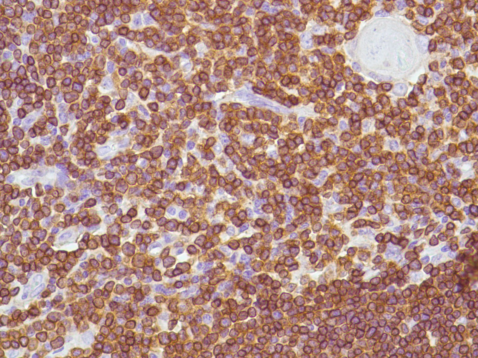
CD20 and CD79a
Species: Dog, Cat (CD20 only)
General information about when this test is indicated:
CD20 is a transmembrane protein found on B-lymphocytes, which plays a role in their differentiation into plasma cells. CD79a is part of the B-cell receptor, the critical part of B-cells responsible for recognising antigens. Therefore, this antibody recognises lymphocytes of B-cell lineage (including plasma cells).
These antibodies are used to confirm and characterise diagnoses of B-cell lymphoma, and to help rule out lymphoma in cases of poorly differentiated “round cell” tumours. They are typically used in conjunction with the T-cell antibody CD3. Some plasma cell tumours are also CD79a positive.
This is helpful because the prognosis and treatment for different forms of lymphoma and plasma cell neoplasia varies according to their classification. There are low-grade T- and B-cell lymphomas that may have a fair to good prognosis, medium-grade B-cell lymphomas that are often chemoresponsive, and high-grade T-cell lymphomas that may be poorly chemoresponsive and may have a poor prognosis. Plasma cell tumours tend to be benign and have a good prognosis compared to some other round cell tumours.
CD31
Species: Dog
General information about when this test is indicated:
CD31 (also known as PECAM-1) is a glycoprotein expressed strongly by endothelial cells, megakaryocytes and platelets; it can also be found on haemopoietic stem cells and leukocytes. It is used during transendothelial migration of leukocytes.
This antibody is useful to identify spindle cells as endothelial, allowing the distinction of haemangiosarcomas, angiosarcomas and lymphangiosarcomas from other “spindle cell” tumours, (e.g. spindloid melanomas and soft tissue sarcomas). This is helpful since malignant vascular tumours often have a worse prognosis (e.g. early metastasis) than soft tissue sarcomas.
c-Kit / CD117
Species: Dog
General information about when this test is indicated:
c-Kit/CD117 is a tyrosine kinase receptor for stem cell factor, found on mast cells, interstitial cells of Cajal and some other cells (e.g. bone marrow stem cells, melanocytes, basal cells of the skin, melanocytes, and germ cells). It is therefore not specific to mast cells, however mast cell tumours generally stain well with antibodies to this receptor and this test is helpful to confirm that diagnosis.
In the intestinal tract, Gastrointestinal Stromal Tumours (arising from interstitial cells of Cajal) are differentiated from other “spindle cell” tumours (e.g. leiomyosarcoma) by this antibody. Many canine GIST’s have aggressive behaviour and may metastasise to local lymph nodes, the mesentery and liver.
Confirmation of a diagnosis of mast cell tumour enables the use of specific treatments such as tyrosine kinase inhibitors. These block signals stimulating cell proliferation from c-Kit and other tyrosine kinase receptors. Constitutive activation of c-Kit is seen in a percentage of canine mast cell tumours in association with mutations in the c-Kit receptor.
Cytokeratin AE1/AE3 and Cam 5.2 (Cytokeratin 8/18)
Species: Dog
General information about when this test is indicated:
Cytokeratins form part of the cytoskeleton in epithelial cells, and less commonly in other cell types. They are found in different combinations in different types of epithelial cells, and therefore specific cytokeratin antibodies can identify these cell lines. Cytokeratin AE1/AE3 is a “cocktail” of antibodies recognising most epithelial cells. Cytokeratin 8/18 identifies most carcinomas, but is not found in squamous cell carcinoma or mesothelioma and so can help to diagnose these.
These antibodies are useful to differentiate carcinomas from each other and other tumour types, which may have a different prognosis and treatment regime. Cytokeratin AE1/AE3 is often used in combination with vimentin.
Ki67
Species: Dog
General information about when this test is indicated:
Ki67 is a nuclear protein that is expressed during all stages of the cell cycle, except G0 (i.e. “resting”, non-dividing cells). Since mitotic figures are sometimes hard to distinguish histologically from artefactual nuclear distortion or necrotic/apoptotic nuclear changes, antibodies to Ki67 allow more sensitive detection of proliferative activity in a population of cells.
The percentage of positive cells produces a quantitative assessment of tumour proliferation. This is useful in tumours that are difficult to prognosticate by histopathological features, such as mast cell tumours and melanomas.
Melan A
Species: Dog
General information about when this test is indicated:
Melan A is a melanocytic antigen. It is very specific for melanomas, but can also cross-react with steroid hormone-producing cells.
This antibody allows the distinction of melanomas from other “round”, “epithelioid” or “spindle cell” tumours (e.g. histiocytic sarcoma, carcinoma, and soft tissue sarcoma). This is useful because the prognosis for malignant melanoma is often more guarded than for some differential diagnoses; and also because specific treatment for melanoma (the “melanoma vaccine”) may be useful in some melanoma patients.
Smooth Muscle Actin
Species: Dog
General information about when this test is indicated:
Smooth muscle actin is part of the contractile apparatus and cytoskeleton of smooth muscle, myofibroblasts, pericytes, liver peri-sinusoidal cells and myoepithelial cells.
This antibody can be used to determine if a tumour or cell of interest arises from smooth muscle, and is used to help diagnose muscle tumours (such as leiomyosarcomas) and to sub-classify mammary tumours. This is clinically useful since spindloid muscle tumours can be indistinguishable from other “spindle cell” tumours (e.g. some forms of melanoma or GIST), and sub-classification of mammary tumours allows better prognostication.
Synaptophysin
Species: Dog
General information about when this test is indicated:
Synaptophysin is a component of the neuronal synaptic vesicle and neurosecretory granules. Antibodies to synaptophysin label neurons and neuroendocrine cells.
This antibody is mainly used to distinguish neuroendocrine tumours from other epithelial or mesenchymal tumours. Neuroendocrine carcinomas can be aggressive and may also secrete hormones.
TTF-1
Species: Dog
General information about when this test is indicated:
TTF-1 is a transcription factor regulating thyroid-specific genes, and also regulating transcription in the brain and lung (Clara cells and Type 2 pneumocytes). TTF-1 antibodies stain most follicular thyroid tumours, some C-cell tumours and some pulmonary carcinomas.
This is useful to differentiate thyroid carcinomas from other epithelial tumours (especially in small biopsies), and to differentiate pulmonary carcinomas from other lesions, including tumours that might have metastasised to the lung.
Vimentin
Species: Dog
General information about when this test is indicated:
Vimentin is a part of the cytoskeleton in mesenchymal cells and mesoderm-derived epithelia (e.g. endometrium and ovary).
This test is used to help distinguish mesenchymal tumours from epithelial tumours, and to identify tumours co-expressing vimentin and cytokeratin (e.g. mesotheliomas).