
JULIE TOMLINSON
Clinical history:
An 18-month old female domestic shorthair cat presented with a bloated abdomen and voluminous mucoid malodorous diarrhoea.
Laboratory results:
CBC revealed a low-normal haematocrit and a marked inflammatory leukogram (see Table 1).
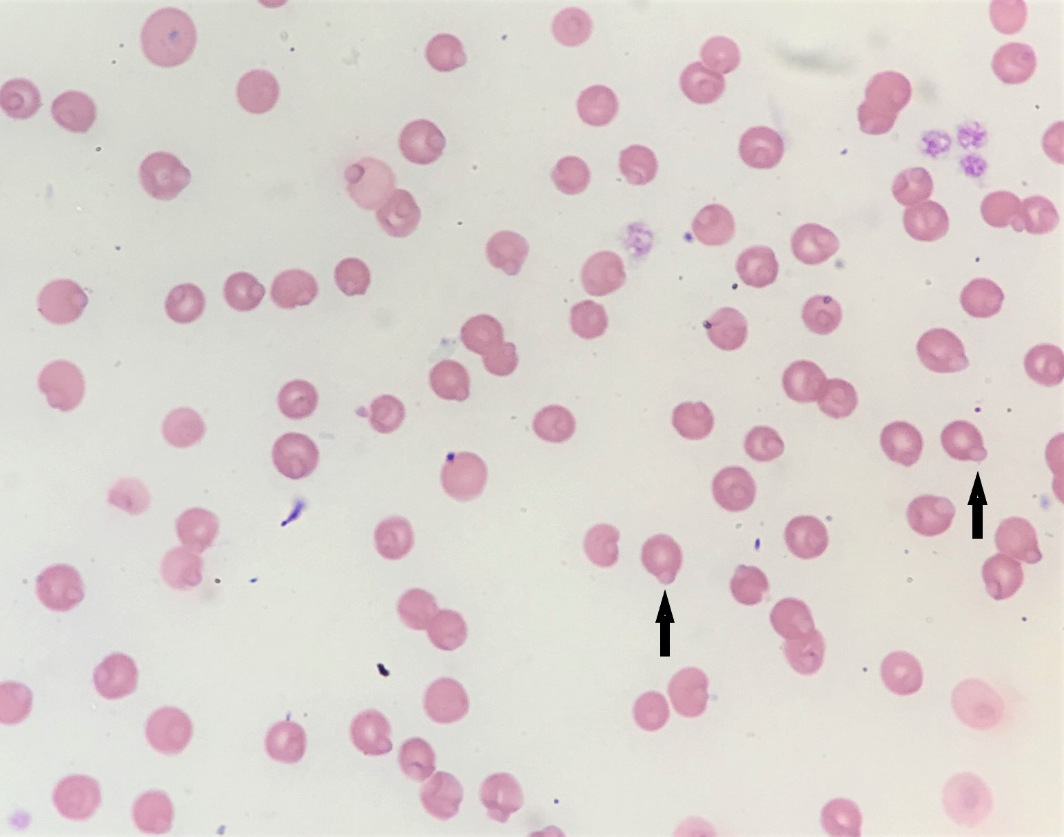
On examination of the blood film, the majority of red cells (approximately 99%) were found to contain Heinz bodies (see Figure 1), which were even more evident when stained with new methylene blue (see Figure 2).
| Table 1. Relevant CBC results | Result | Reference interval |
| HCT / PCV | 0.26 | 0.24-0.45 L/L |
| Reticulocytes absolute | 37.32 | 0-50 x 109/L |
| WBC | 71.1 H | 7-20 x 109/L |
| Neutrophils absolute | 56.2 H | 2.5-12.5 x 10⁹/L |
| Bands absolute | 7.1 H | 0-0.3 x 10⁹/L |
| Lymphocytes absolute | 5.7 | 1.5-7 x 10⁹/L |
| Eosinophils absolute | 2.1 H | 0-1.5 x 10⁹/L |
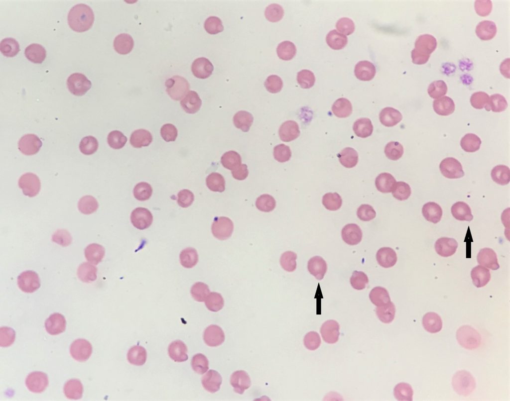
Figure 1: Blood smear, Wright’s stain 100x. Arrows – Heinz bodies.
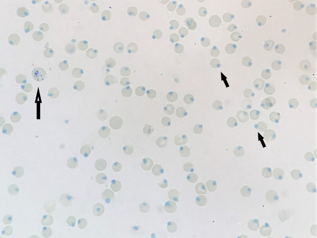
Figure 2: Blood smear, New Methylene Blue stain 100x. Large arrow – aggregate reticulocyte. Small arrows – Heinz bodies.
Diagnosis:
Differentials to consider included acute Heinz body haemolytic anaemia – commonly seen with ingestion of exogenous oxidants such as paracetamol, onions, garlic, etc. Although the HCT was technically within the reference interval, it was low normal which could reflect an anaemia for this individual. There was no evidence of reticulocytosis to suggest regeneration, but excluding acute pregenerative hemolysis would require trending the CBC as it can take 2-3 days to see a reticulocytosis.
Large numbers of “endogenous” Heinz bodies in the absence of haemolysis have also been associated with conditions including diabetes, hyperthyroidism, and GI lymphoma. Although many of these cats are not anaemic, they may have shorter RBC lifespans and lower haematocrits than cats with the same conditions that lack Heinz bodies.
Unfortunately, the cat in this case was euthanized due to financial constraints and the concern for clinical deterioration. No additional testing was performed thus the underlying cause for the oxidative damage was undetermined. GI lymphoma was a consideration given the clinical history of diarrhoea. Differentials to consider for the marked inflammatory leukogram would include GI perforation/septic effusion or possibly a paraneoplastic leukocytosis. Although we can see a marked inflammatory leukogram with haemolysis as a result of IMHA, I would not expect such a marked inflammatory response with Heinz body haemolytic anaemia.
MANY THANKS TO NICOLE ROBERTSON FROM PET DOCTORS, PAPATOETOE FOR SUBMITTING THIS CASE