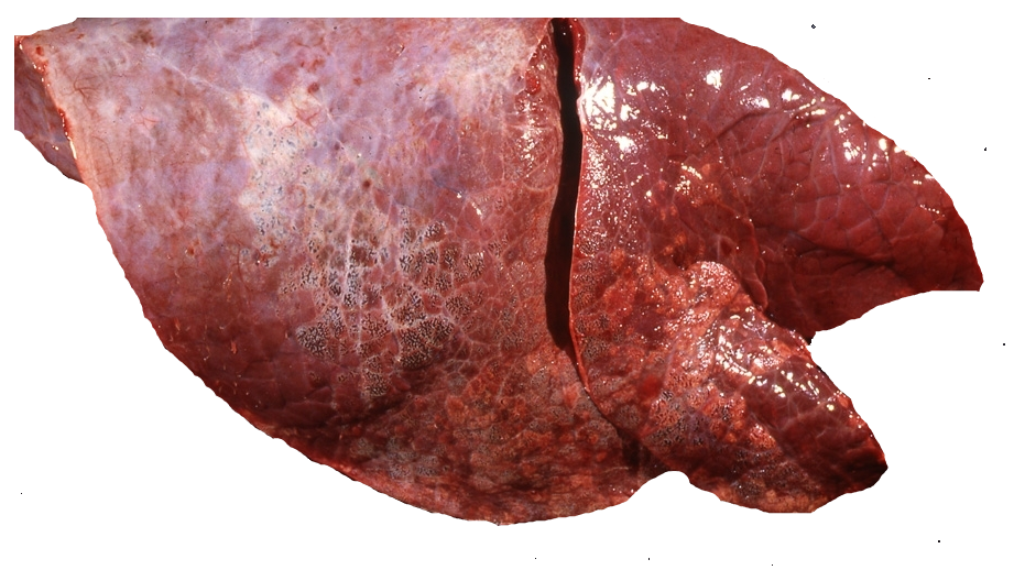
Here are two photos courtesy of Keith Thompson (Massey University) from a cow that died. What is your diagnosis?
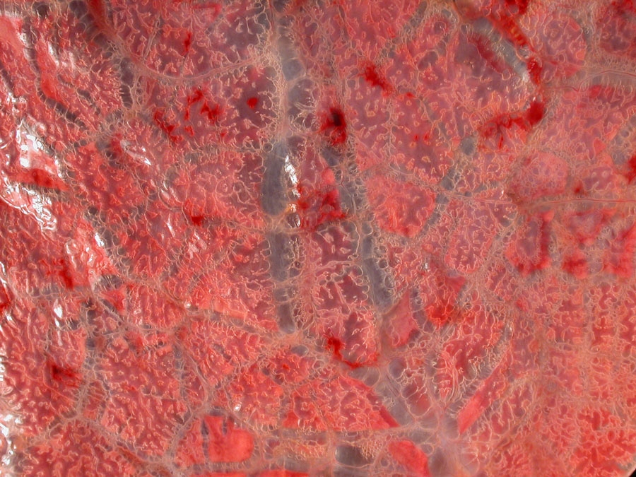
Diagnosis: Interlobular and subpleural emphysema (severe).
Discussion: This is a common finding in cows and it is usually not as severe as this. It is the result of exaggerated or gasping respiration (with a closed glottis) leading to rupture of alveoli. It is a secondary lesion. What many do is see emphysema like this and assume they are dealing with acute bovine pulmonary edema and emphysema (or that really awful term “fog fever” which has zilch to do with fog). Of course, emphysema can be seen in that entity but it is not exclusive to it – any disease that causes exaggerated or gasping respiration can lead to this.
Now, have another look at the above photos and notice that the lung tissue is nice and pink, it is normal. There is no underlying respiratory disease in this case.
Although this type of emphysema is a secondary change it can still be very important in animals that survive. You might have a downer cow from milk fever, for example, that develops severe emphysema. You successfully treat the milk fever but the cow remains depressed and ends up being put down. It is the emphysema that is the cause of this.
What might be less well known is that in some cows the air may further escape into the mediastinum and then through the thoracic inlet where it makes its way into the dorsal subcutaneous tissues. Many of you have felt those cows with crackling under the dorsal skin. Again, a secondary change but a very important and life-limiting secondary change.
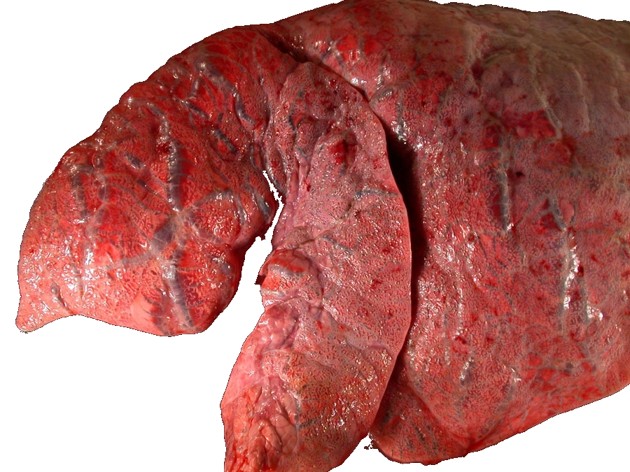
Photograph 3 (below) is from Ryan Luckman at Waimate Veterinary Centre. I love getting photos and Ryan’s great photo nicely shows severe emphysema. Note too, the normal light pink lung colour. Carla Fletcher from Gateway has also sent me photos of a similarly good case.
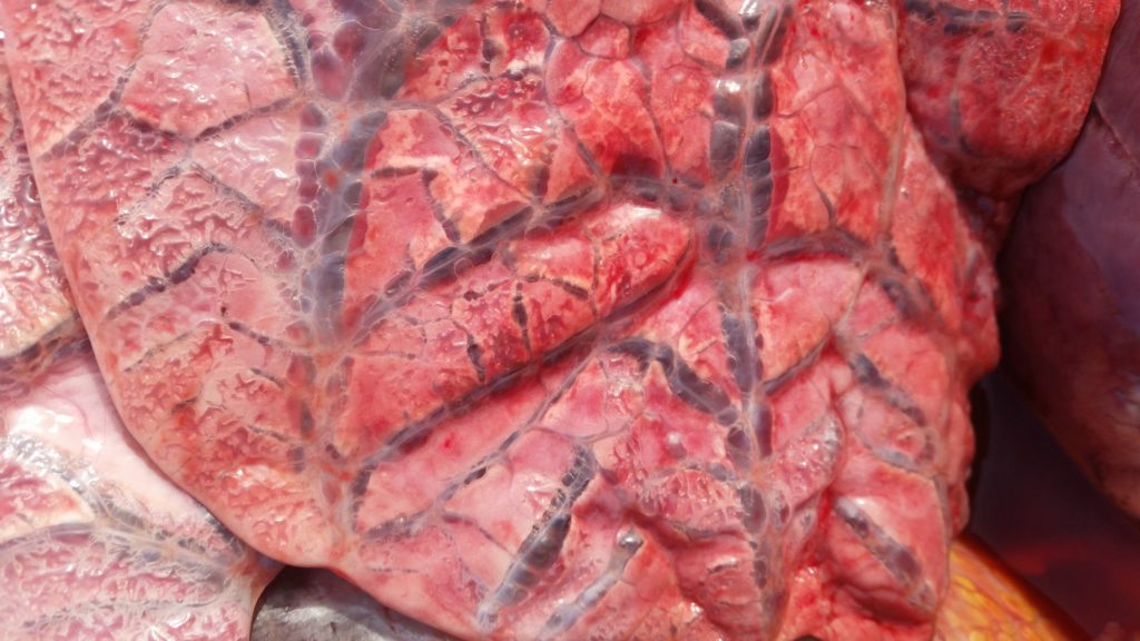
Photograph 4 (below) is a case where there is an underlying lung disease – the lung in this photo from Keith Thompson has a solid appearance (this is from a case of acute bovine pulmonary edema and emphysema).