


SANDY WELTAN
This is a case I encountered in South Africa, but considering the high incidence of Johne’s disease in New Zealand, I thought it would be of interest and possibly relevant here too.
Clinical history:
A 2-year-old, neutered, male Dachshund (right) presented to his veterinarian with lethargy inappetance, mild pyrexia and abdominal pain. Enlarged mesenteric lymph nodes were found on abdominal ultrasound examination.
Laboratory testing:
Cytology of mesenteric lymph node, spleen and sternum revealed numerous macrophages containing vast numbers of negatively staining bacterial rods (Figure 1).
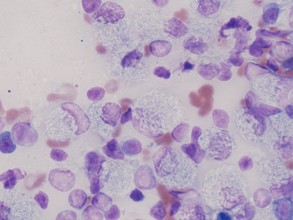
Lymph node biopsies were sent to The Centre of Excellence for Biomedical Tuberculosis Research at Stellenbosch University in Cape Town, South Africa. Following culture, PCR and sequencing, the bacteria were identified as Mycobacterium paratuberculosis.
Treatment:
Treatment was started with rifampicin clarithromycin, doxycycline and supportive therapy. There was a temporary clinical response but bacteria were still demonstrated in smears from lymph nodes. Despite more aggressive therapy, his condition deteriorated and he was euthanased. A post-mortem was performed.
Post-mortem results:
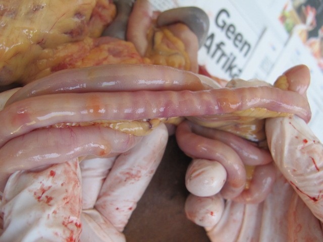
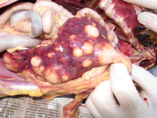
Nodular lesions were found in the intestine, spleen and kidney (Figures 2 and 3). Ziehl-Neelsen staining of histopathology slides revealed epithelioid macrophages and Langerhan’s giant cells engorged with acid-fast filamentous bacteria.
Although we were unable to confirm the source of infection, the dog’s history included exposure to sheep farms in the Western Cape region. This report demonstrates that MAP infection should be included as a potential cause of gastrointestinal disease in companion animals, especially those that may be exposed to premises with infected livestock.