
GEOFF ORBELL
Clinical history:
Fourteen rising 1-year-old beef bulls and steers died suddenly over a 24-hour period five days after being yarded for drenching (pour-on) and castration. The cattle had been bought up from the South Island (in February) for finishing and had reportedly been previously vaccinated twice for clostridial disease prior to transport. Both castrated and uncastrated cattle were dead.
Pathology:
On post-mortem examination of multiple cattle, the subcutaneous tissue on the ventrum was oedematous and yellow, and extended into underlying muscle in both castrated and non-castrated animals. Clinically Blackleg was suspected and post-mortem samples were submitted for culture and histology. Histologically the lesions were more consistent with malignant oedema with the subcutaneous tissue and fascia primarily affected. Anaerobic culture failed to isolate any clostridia.
Treatment and further investigation:
All remaining cattle were treated with high dose penicillin and vaccinated for clostridial disease. Another five died over the next three days with no more deaths five days after vaccination.
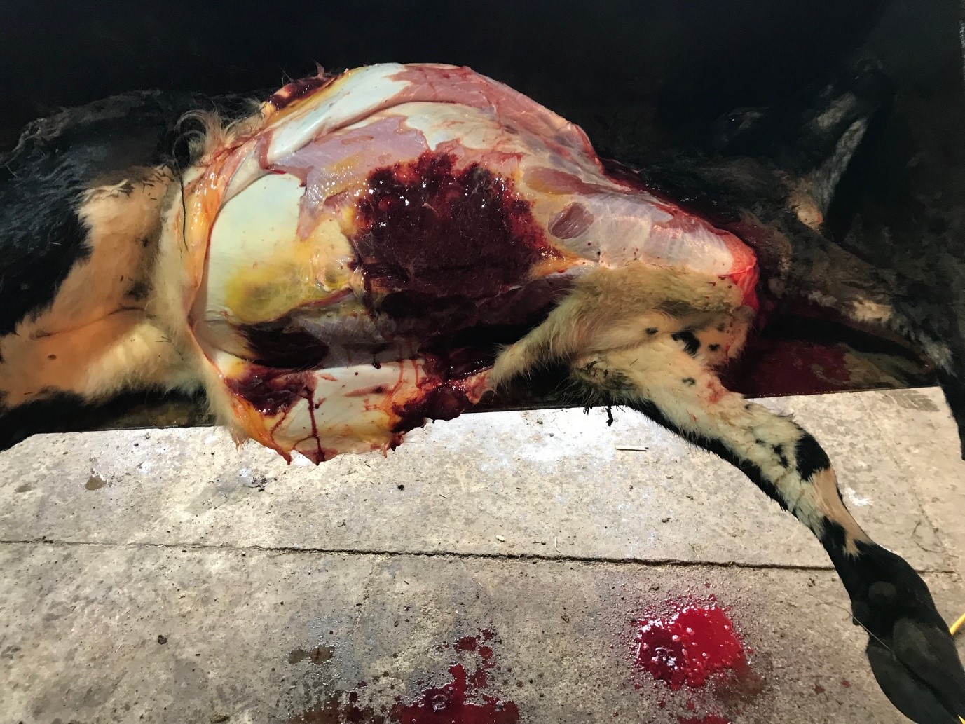
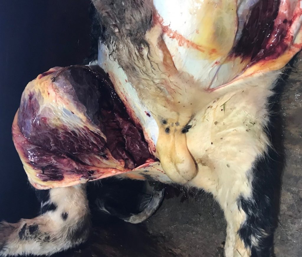
One bull was lame with a markedly swollen upper right hind-limb, but was systemically well. The decision was made to euthanase this animal and submit further samples for histology and culture. On post-mortem examination there were multiple areas of yellow, oedematous subcutaneous tissue on the ventrum, poll, distal limbs and groin. The proximal hind-limb muscles were markedly swollen, darkly discoloured and haemorrhagic.
Histologically there were similar oedematous changes to the first post-mortem but no clostridia were identified and there were more acute and chronic lesions involving the hind-limb muscles. Anaerobic culture was unrewarding from the affected muscle.
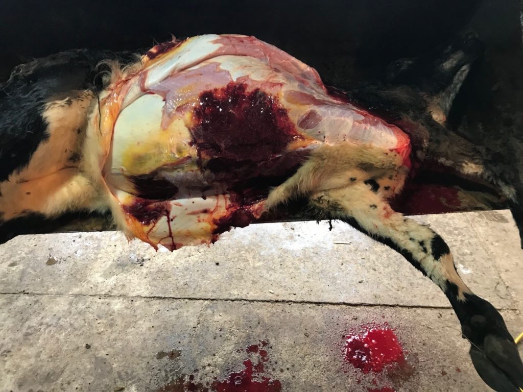
Post mortem examination of the euthanased bull demonstrating extensive muscle haemorrhage and necrosis and subcutaneous oedema.
Discussion:
These cases are most consistent with Malignant Oedema but Blackleg could also be involved, which would explain the sudden death of uncastrated cattle and the changes in the thigh muscles of the euthanased bull. Tissues from both cattle have been sent overseas for immunohistochemistry to help identify the clostridial species involved.
Although Malignant Oedema and Blackleg are both clostridial diseases, the disease pathogeneses and usually the pathogens involved are different, although Clostridium chauvoei can cause both.
Malignant Oedema usually occurs secondary to soil contamination of wounds, in this case castration wounds, whereas Blackleg is caused by sporulation of pre-existing clostridial spores in the muscle due to creation of a localised anaerobic environment (usually blunt trauma e.g. yarding).
Malignant Oedema has been associated with Clostridium septicum, C. sordellii, C. chauvoei, C. novyi and C. perfringens. Blackleg is only caused by C. chauvoei. It is worth noting that Malignant Oedema is the only disease in New Zealand ruminants where C. sordellii has been confirmed to cause clinical disease.
Vaccination in the face of an outbreak is often effective in reducing the morbidity and mortality rate, although deaths will usually continue for a number of days until there is an adequate immune response. A booster vaccine is still recommended 2-4 weeks later even if mortalities have ceased. Live clinically affected stock are rarely seen with both Malignant Oedema or Blackleg, and very rarely saved, but in some cases high dose penicillin can be effective in preventing mortality but clinical lesions will still persist in affected tissue reducing carcass value.
Vaccination for clostridial disease is usually extremely effective in prevention when label directions are followed and boosters are given. In this case, disease could have been due to lack of vaccination, improperly handled/administered vaccine, or less likely vaccine failure.
This case is therefore unusual, but the previously reported vaccination was not able to be confirmed through correspondence with the previous owner.
Diagnosis of cases of suspect Blackleg or Malignant Oedema usually requires histology or culture, but these are often adversely affected by autolysis caused by post-mortem clostridial proliferation. Culture from dead cases of suspect Malignant Oedema is more complicated, in that some of the causative clostridia are also normal post-mortem invaders, e.g. C. septicum and C. sordellii.
Ideally fresh samples should be from recently dead stock and include large pieces of affected muscle or oedematous fascia, so that anaerobic sampling can be taken from the centre of affected tissue, as exposure to oxygen greatly recues the chance of successful culture. Histology should be performed on multiple smaller samples (1cm x 2cm x 1cm) from affected muscles and fascia to ensure rapid fixation.
Thank you to Cosmin Susa from Hunterville Vet Clinic for providing the images and clinical feedback on this case.