
LISA HULME-MOIR
Many of us are familiar with the classic cases of young bull terrier type dogs coming in with demodicosis. However not many may have Demodex at the forefront of our minds when working up skin disease in older animals.
A recent case highlights the value of skin cytology and the need to keep demodicosis on the differential list for older dogs, especially those on immunosuppressive therapy.
Clinical history:
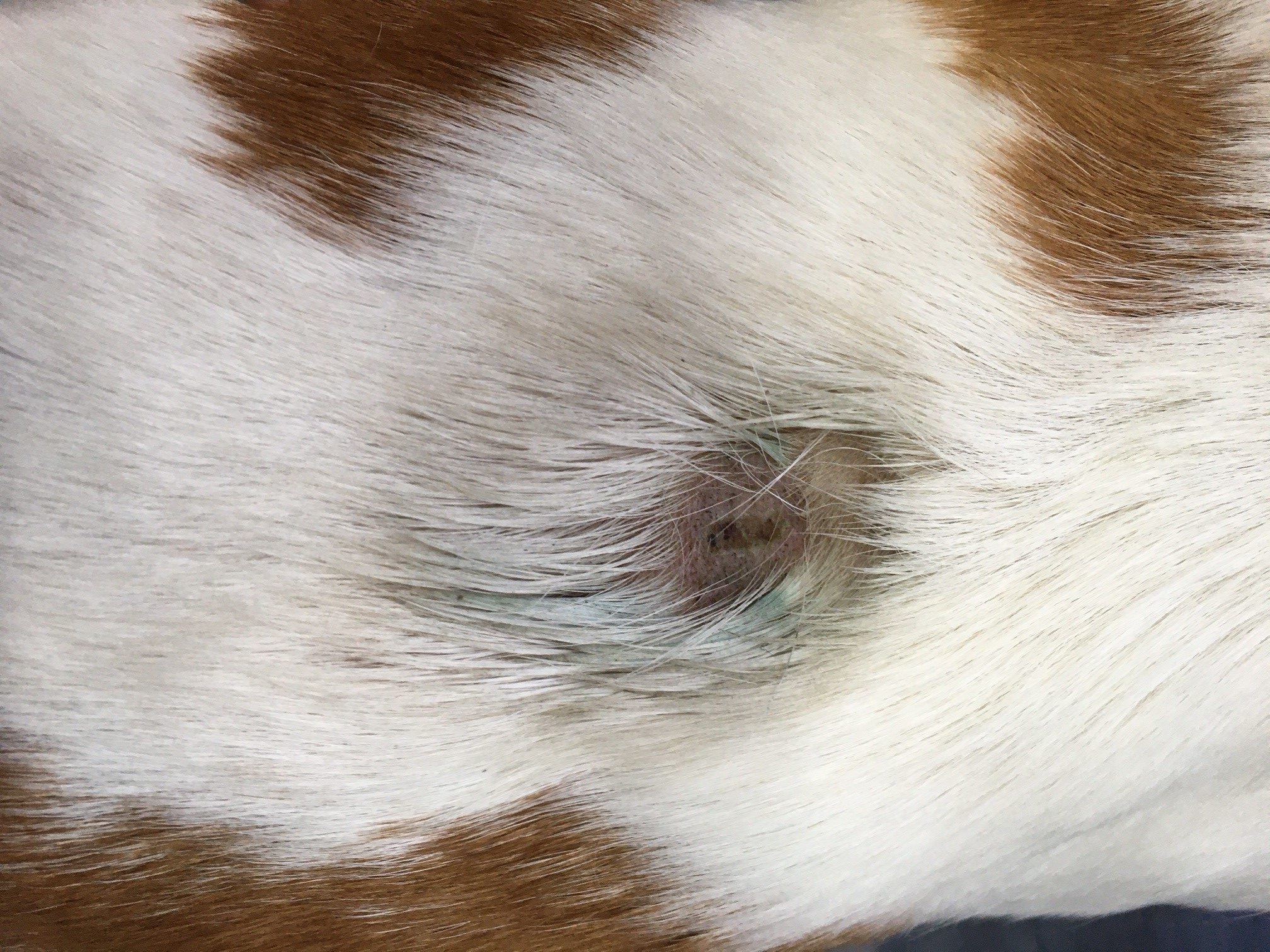
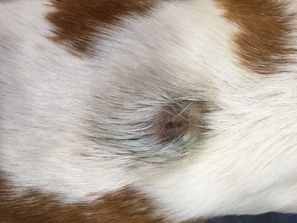
Skin scrapes and tape preparations were received from a middle-aged, female Bassett hound, who had a hot-spot on the mid-dorsal back (Figure 1). She had a history of atopic dermatitis, which was currently being well-controlled with oclacitinib.
Cytology:
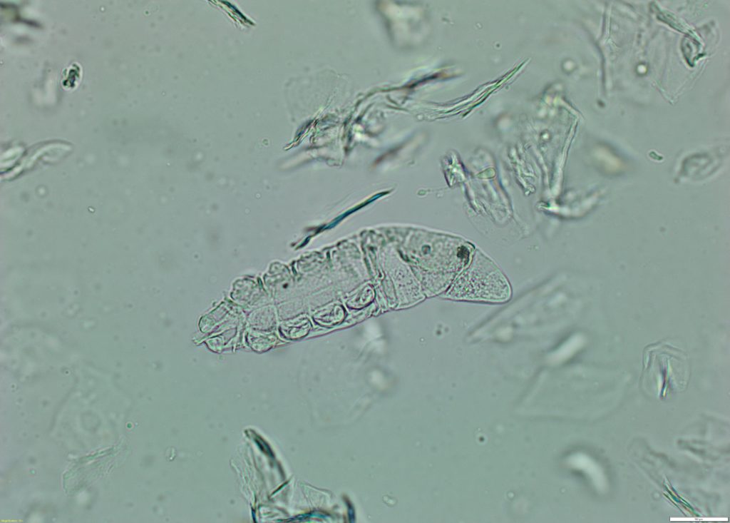
Suppurative inflammation with intracellular bacterial cocci confirmed the presence of a bacterial pyoderma. Additionally, many Demodex mites, up to 10 per slide, were seen (Figure 2).
Discussion:
In a recent consensus statement issued by the World Association for Veterinary Dermatology, the finding of more than one Demodex is considered indicative of clinically relevant demodicosis.1
Many dogs with adult onset demodicosis have an underlying cause of immunosuppression identified. Common underlying causes include hyperadrenocorticism, hypothyroidism, leishmaniasis (not present in New Zealand), T-zone lymphoma and other neoplasms, corticosteroids and other immunosuppressive drugs including chemotherapy.2,3 In this case, it is likely that the oclacitinib contributed to the proliferation of the mites. This risk is listed as a precaution on the prescribing information for this product. Secondary bacterial infection is a common sequelae of demodicosis.
It has been suggested in some quarters that we may start to see less demodicosis with the rise in use of isoxazolines for routine parasite control. These have been found to be highly effective in controlling Demodex.1 On further enquiries with the present case, the owners had been using a supermarket brand of parasite control. The dog had an excellent response to treatment of the lesion with a topical anti-inflammatory and antimicrobial preparation, plus institution of routine parasite control with an isoxazoline product.