
BERNIE VAATSTRA
Clinical history:
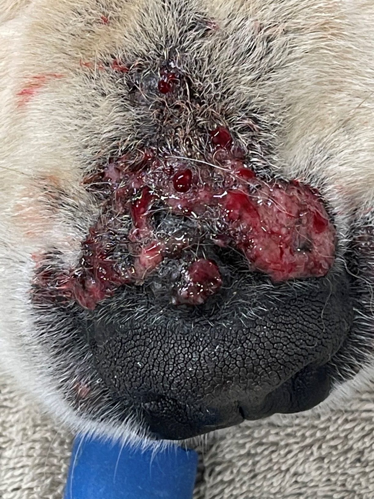
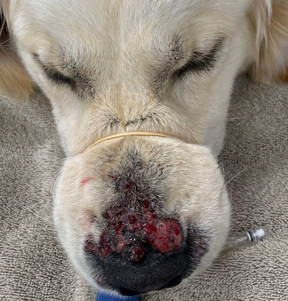
A one-year-old, male, neutered Golden Retriever developed erupting, ulcerated, erythematous and crusting lesions over the dorsal surface of the nasal planum (Figures 1 and 2). The lesions developed quickly over approximately 48 hours, accompanied by vomiting and diarrhoea.
Clinical differentials included bacterial furunculosis, fungal or yeast infection, tick-bite hypersensitivity, facially oriented autoimmune disease, and eosinophilic furunculosis.
Skin biopsy samples were collected for histology and culture.

Laboratory testing:
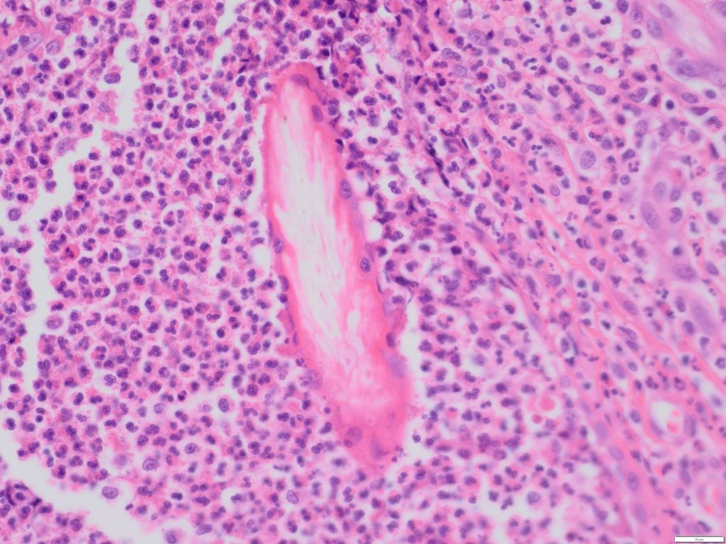
Histological examination revealed extensive epidermal ulceration and crusts of necrotic debris. Follicles were disrupted by large numbers of eosinophils and fewer macrophages, neutrophils and lymphoid cells. Similar inflammatory cells surrounded free hair shafts within the perifollicular dermis (Figure 3). A few Gram-positive cocci were observed in the surface crusts of Gram-stained sections. No fungal elements were detected on PAS sections.
Culture of the fresh tissue produced a heavy growth of Staphylococcus intermedius group. The bacteria were sensitivity to amoxicillin-clavulanic acid, cephalothin, enrofloxacin, tetracycline, and trimethoprim-sulpha and resistant to penicillin (based on disk diffusion testing).
Diagnosis:
Eosinophilic furunculosis of the face with secondary staphylococcal infection.
Discussion:
Canine eosinophilic furunculosis is an uncommon dermatopathy with an alarmingly sudden onset. Young, large breed, inquisitive dogs are overrepresented, with lesions typically occurring on the face and occasionally on other extremities. While the exact aetiology is unknown, the highly eosinophilic nature of the lesions and predilection for adventurous dogs, strongly suggests a reaction to arthropod or insect bites. In some cases, there is documented interaction with bees, wasps or ants. Lesions are characteristically eruptive, exudative, crusted and pruritic. Some dogs develop fever, malaise, anorexia, and vomiting or diarrhoea.
Fortunately, the prognosis is very good with the majority of dogs responding rapidly to systemic glucocorticoids.
In the present case, a short course of prednisone and Clavulox® resulted in complete resolution of the lesions apart from a small scab by the time of follow up (approximately10 days after presentation). A satisfying outcome for all concerned!
Many thanks to Robert O’Connor of New Plymouth Veterinary Group for the case submission and excellent gross photographs.