Clinical history:
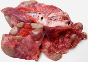
A mob of 200 weaner deer were transferred between properties the same day as being given a mineral and anthelmentic drench. They were in good health on arrival at the new farm, but a few days after being fed silage they started dying. Most of the mob died over a 4-5 day period. Multiple lesions were seen in the lung with suspect lungworm (see photo right).
Lung was sent to the laboratory for histology testing, along with intestinal contents for enteric screen, faeces for faecal egg count, and liver for trace element testing.
Laboratory testing:
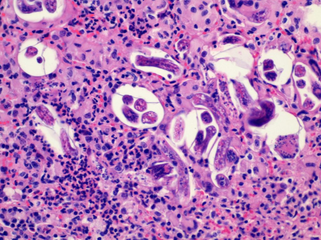
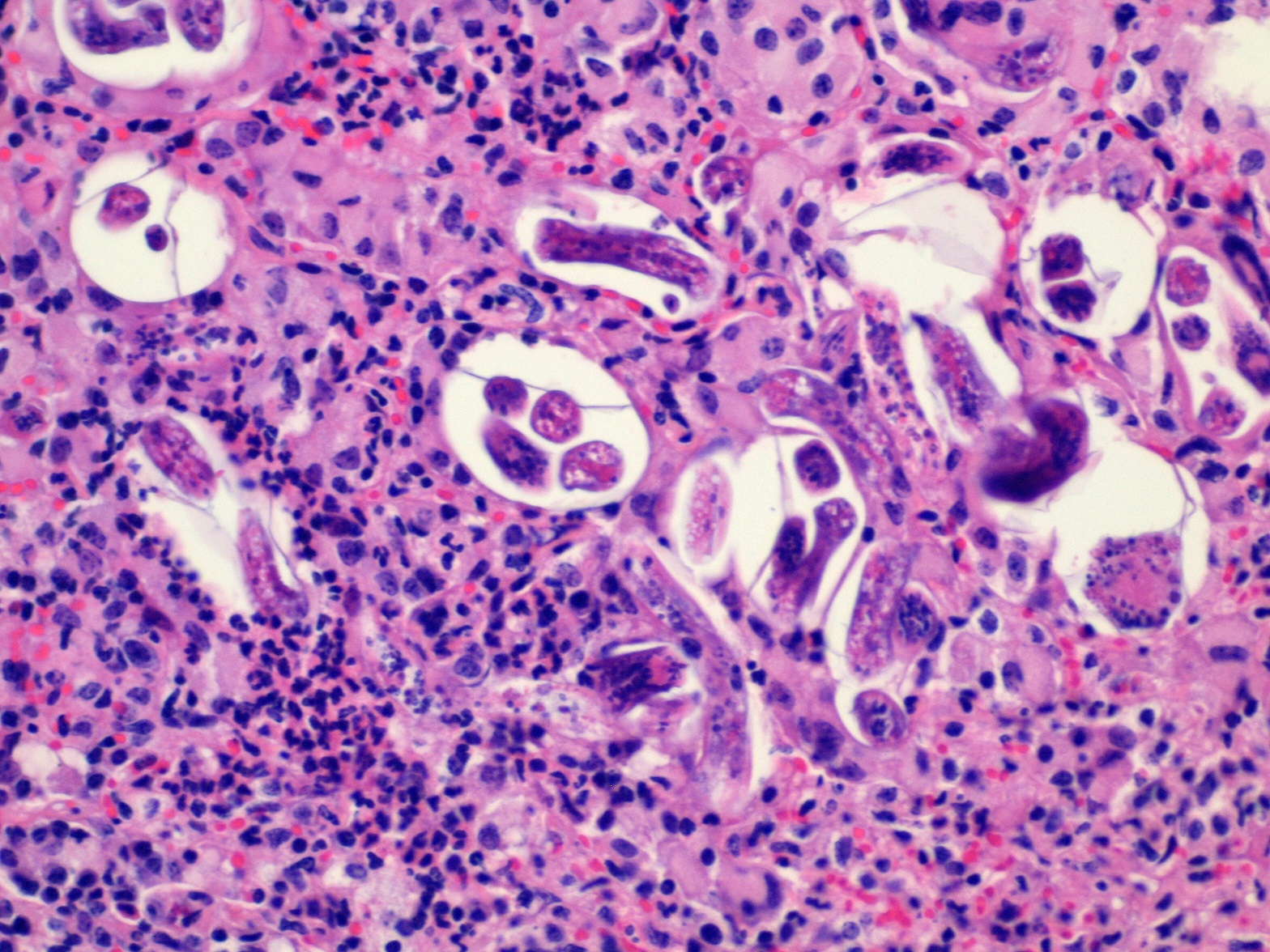
Intestinal contents grew Yersinia enterocolitica; copper and selenium levels were within the adequate range; 600 strongyle eggs/g were seen in the faeces. Histology of the lung revealed many airways containing lungworm larvae admixed with eosinophils, neutrophils, and karyorrhectic debris. Adult nematodes were present with large airways. There was marked bronchial mucosal hyperplasia. Moderate numbers of lymphocytes, plasma cells, and eosinophils were found to expand the interstitium. Alveoli were often enlarged and coalesced.
Diagnosis and comments:
Histology confirmed severe lungworm infestation in the deer. The lesions were serious enough to explain respiratory failure and death.
Weaner deer in their first autumn and winter are the most susceptible to infection. This is because they have not yet developed full immunity and lungworm larvae are often present in high numbers during this time. Deer of all age groups may carry burdens but usually young stock or heavily stressed animals will develop more severe infections. Moving naïve animals onto an infected property may have triggered an outbreak in this case.
Photograph below: Lungworm larvae in alveoli with associated inflammation. H&E 200x.