

AMY WEEDEN
Clinical history:
An adult, castrated male, Oriental breed cat presented with severe diarrhoea and cachexia. Abdominal ultrasound revealed several abnormalities to include mild diffuse small intestinal changes, moderate hepatopathy/hepatomegaly, bilateral chronic renal degeneration, and trace peritoneal effusion.
Laboratory results:
Serum biochemistry identified azotaemia and hyperbilirubinaemia with a moderate non-regenerative anaemia and an inflammatory leukogram on the CBC. The cat was also likely FIV positive.
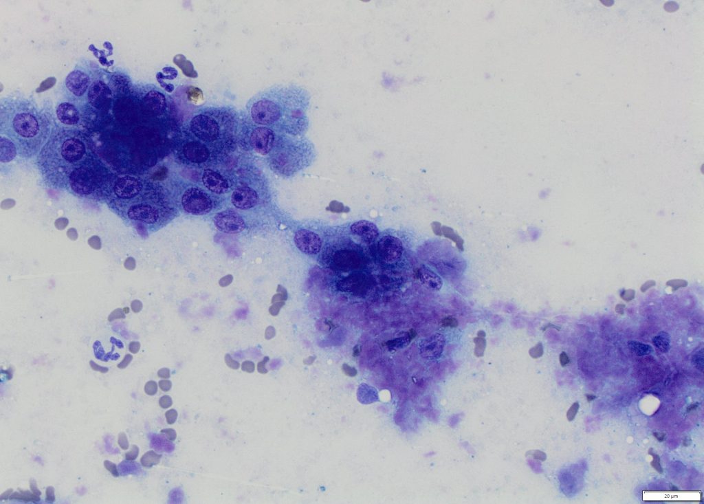
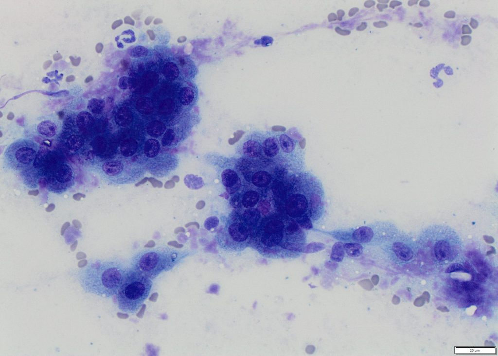
Hepatic aspirate and peritoneal effusion cytology were performed. The effusion was most consistent with a high-protein, modified transudate.
Diagnosis:
Hepatic amyloidosis with moderate neutrophilic inflammation and likely hepatocellular hyperplasia.
Discussion:
The most notable cytologic finding in this case is the pink proteinaceous material surrounding the hepatocytes. This is an infrequent cytologic finding, which is highly suggestive of amyloid. For confirmation of amyloid, a Congo-red stain was performed. The proteinaceous material stained red-orange with birefringence observed on polarized light. This confirmed that the material was amyloid. Renal samples were not submitted, but the presence of renal azotaemia with significant proteinuria suggests that renal amyloidosis was also present.
Amyloidosis is not a specific disease. Rather it refers to a group of protein misfolding disorders characterized by deposition of amyloid protein in various organs and tissues. AA-amyloidosis is the most common form of amyloidosis in domestic animals.1 Amyloid protein AA is an amino-terminal fragment of the acute phase protein serum amyloid A or SAA.2 SAA is produced in response to tissue injury associated with chronic inflammatory or neoplastic diseases, or amyloid deposition can be idiopathic.1,2 Clinical presentations will vary, based on the organs affected and the severity of the amyloid deposition. Reactive systemic amyloidosis secondary to chronic inflammatory conditions may affect liver, kidneys, spleen, lymph nodes, and adrenal glands. The kidney is the main target organ for the deposition of amyloid in familial amyloidosis in Abyssinian cats and Shar-Pei dogs, and the liver tends to be the target organ in Siamese cats.1
Hepatic amyloidosis is uncommonly reported in cats, and the majority of cases are either Siamese or Oriental breeds. Prognosis is usually poor with most of the reported cats either being euthanized or dying acutely; hepatic rupture with haemoabdomen is reported in cats affected with this condition.3 According to one study, cats with FIV are more likely than the general cat population to have amyloid deposits in their tissues,4 and that may have been a predisposing condition in this case, in addition to breed predilection.
Thank you to Tommy Fluen and Veterinary Specialist Group in Auckland for this interesting case.
References:
1. Woldemeskel M. A concise review of amyloidosis in animals. Vet Med Int. 2012:427296, 2012.
2. Nelson RW and Couto GC. Glomerulonephropathies in Small Animal Internal Medicine, ed 3, Mosby, p.602, 2003.
3. Neo-Suzuki S, Mineshige T, Kamiie J, et al. Hepatic AA amyloidosis in a cat: cytologic and histologic identification of AA amyloid in macrophages. Vet Clin Pathol. 46:331-336, 2017.
4. Asproni P, Abramo F, Millanta F, Lorenzi D, Poli A. Amyloidosis in association with spontaneous feline immunodeficiency virus infection. J Feline Med Surg. 15:300-306, 2013.