
LISA HULME-MOIR
Clinical history:
A 15-year old male, neutered Chihuahua presented with a large 6x5x3cm subcutaneous mass in the left inguinal region. A fine needle aspirate of the mass was submitted to the laboratory for cytology.
Laboratory results:
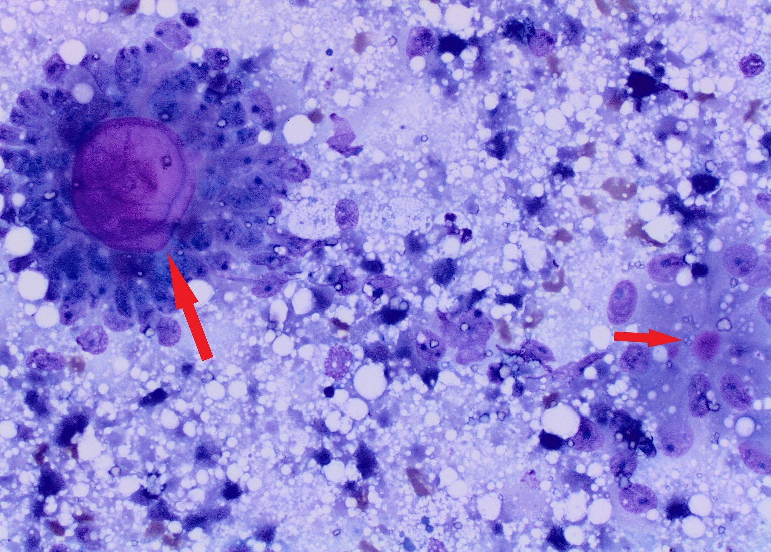
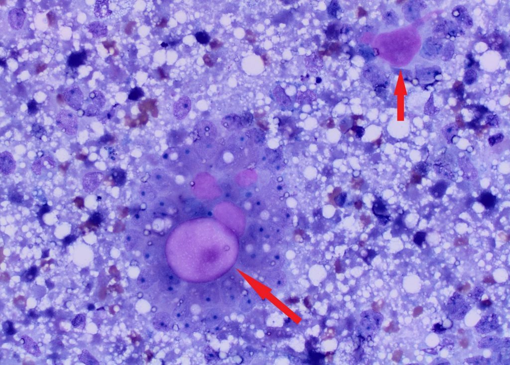
On cytology, the submitted slides contained many medium to large cells that were frequently arranged in rosette fashion around variably sized globules of bright pink hyaline material (photographs 1 and 2). The cells were often poorly preserved but when intact appeared round to columnar in shape with large amounts of pale blue cytoplasm, often with variably sized vacuoles.
Photograph 1 & 2: Fine needle aspirate of an inguinal mass in a dog. Many ill-defined cells are arranged in rosettes around a pink glassy appearing matrix (arrows). Many variably sized, clear vacuoles are present in the background and in the cytoplasm of the few intact cells.

Diagnosis: Sertoli cell tumour with Call-Exner bodies
Discussion: Testicular tumours should be considered as a differential for inguinal masses in male dogs, particularly small breed dogs, which have an increased incidence of cyrptochidism.1 Cryptorchid testicles are predisposed to Sertoli cell tumours and seminomas and also have an increased risk of torsion.2
Cytologically, Sertoli cell tumours are characterised by the trabecular and palisading arrangement of cells and the large vacuoles contained within the cytoplasm.3 The case presented here had a further distinguishing feature of having large variably sized lakes of pink hyaline material around which the cells were arranged. These are termed ‘Call-Exner’ bodies. Call-Exner bodies have been identified in both humans and veterinary species, most often in granulosa cell tumours of the ovaries but also in other sex cord tumours such as gonadoblastomas and Sertoli cell tumours.4 They are believed to be an attempt by the neoplastic cells to form a basement membrane.
Thank you to Glenfield Veterinary Clinic for sending excellent smears and this interesting case.
References:
1. Yates et al. Incidence of cryptorchidism in dogs and cats. Vet Record.152:502-504, 2003
2. Reif et al. A cohort study of canine testicular neoplasia. J Am Vet Med Assoc. 175:719-723, 1979
3. Moreau, AM and Scruggs, JL. What is your diagnosis? Inguinal mass in a dog. Vet Clin Pathol. 47:155-156, 2018
4. Masserdotti et al. Cytological detection of Call-Exner bodies in Sertoli cell tumours from 2 dogs. Vet Clin Pathol. 37:112-114, 2008