

SANDY WELTAN & CRISTINA GANS
Clinical history
Bella, a 10-year-old female entire Shih Tzu presented for evaluation of abdominal distension. Aside from this, Bella was well at home, with no changes in appetite, water intake, urination or defaecation noted. Fourteen months prior, Bella had a cystic mammary lump surgically removed. Unfortunately, histology had not been performed for this mass.
On examination, marked abdominal distension was present. Ultrasound scan demonstrated large volume abdominal effusion, abnormal appearance to both ovaries bilaterally, cystic lesions throughout the uterine wall, and occasional nodular appearance to the peritoneal fat. Three view thoracic radiographs did not show any overt metastatic disease.
Cytology
A sample of the peritoneal fluid was submitted to the laboratory. Direct and cyto-centrifuged smears were made from the fluid.
The cellularity was high, even in the direct smears which contained moderate numbers of erythrocytes and clusters of hyperplastic mesothelial cells, as well as clusters of other atypical cells which were present in small and larger groups. They were loosely cohesive and showed marked anisocytosis and anisokaryosis with a high nucleus to cytoplasm ratio. The cytoplasm was moderately basophilic with irregular cell borders with membranous projections. The nuclei were oval and contained coarse chromatin with large but indistinct nucleoli. (Figure 1).
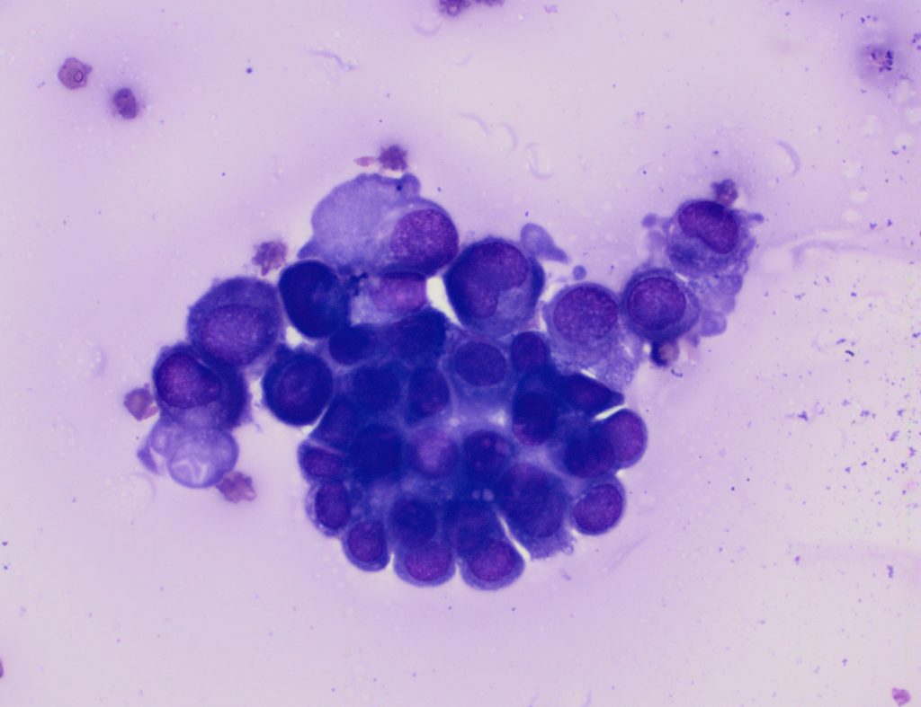
Background cells consisted of lymphocytes, non-degenerate neutrophils and macrophages, many of which were erythrophagocytic and cytophagic. This suggested a neoplastic effusion (possible carcinomatosis).
Histology
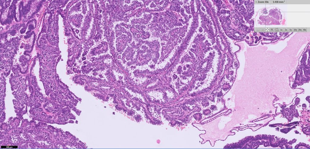
Bella underwent ovariohysterectomy with uterus and ovaries submitted for histology, along with biopsy of nodular peritoneal fat. On histopathological examination, both ovaries displayed an infiltrative neoplasm consistent with an ovarian carcinoma (Figure 2). The presence of neoplastic emboli was evident in the lymphatics in these sections.
The peritoneal fat did not show any evidence of neoplasia. There was cystic endometrial hyperplasia and adenomyosis in the uterus which is a common incidental finding in older intact female dogs.
“Ovarian carcinoma is common in humans, but uncommon in dogs.“
Follow-up
The abdominal effusion completely resolved post-ovariohysterectomy, and to date (five weeks later), there has been no recurrence. Bella’s owner has elected not to pursue any follow up oncology treatments.
Discussion
Bilateral ovarian carcinomas have been reported in dogs, although this is less common than unilateral carcinomas. Several studies have reported regional or localised metastasis (including peritoneal carcinomatosis).
Thanks to Julia Giles and Holly Smith of Totally Vets Manawatu for providing the clinical information for this case.
References:
> Tumors in Domestic Animals. John Wiley & Sons, Inc. Fifth Edition. 2017.
> Patnaik, A. K., and P. G. Greenlee. Canine ovarian neoplasms: a clinicopathologic study of 71 cases, including histology of 12 granulosa cell tumors. Vet Path. 24:509-514, 1987.Sforna M, Brachelente C, Lepri > E, Mechelli L Canine Ovarian Tumours: a retrospective study of 49 cases. Vet Res Commun 27 Suppl 1: 359-361, 2003
> Solano-Gallego, L. and C. Masserdotti, Reproductive System, in Canine and Feline Cytology. R.E. Raskin and D.J. Meyer (eds). A Colour Atlas and Interpretation Guide, Pp. 313-352, Elsevier, St Louis, Missouri, 2016.