
MICHAEL HARDCASTLE
As veterinarians we are trained to describe lesions accurately, but sometimes language can fail, however “a picture is worth a thousand words”. Therefore, the submission of images alongside a written history is always encouraged and welcomed for every case we receive at the laboratory.
When to submit images
The site of biopsies and the selection of lesions to sample can be critical in their interpretation. Images can be very helpful in anchoring our findings around the clinical process, whether it is a skin problem in a dog or lung lesions in a cow. We also find clinical and radiographic images particularly helpful in providing context when interpreting small biopsies of organs or bone lesions. The relationship between biopsy size and lesion size, and the location of the biopsy (i.e. whether or not it would have been representative of the lesion) serve to inform our confidence in a diagnosis and our ability to rule out other differential diagnoses.
Occasionally, we also receive fixed samples that have warped, changed in colour and texture, or fragmented in transit. Surgical margins or the location of the mass/lesion within the tissue may not be obvious once these changes have occurred. Images of the fresh lesion in situ and/or after removal can be helpful in grossing the specimen and deciding where to collect sections to examine areas of concern to the surgeon.
The diagnostic value of images
Case 1
A 17-year-old, Domestic Short-Hair cat underwent multiple skin biopsies since a veterinarian wished to rule out epitheliotropic lymphoma. The initial history provided was that the patient had been treated with monthly injections of methylprednisolone acetate.
Histopathology mainly demonstrated cicatrical alopecia (i.e. fibrosis and loss of hair follicles and adnexa), a non-specific change suggestive of a previous skin injury such as trauma or ischaemia. Acquired skin fragility was mooted as a possible underlying problem given the history of glucocorticoid therapy.
Further discussion with the treating veterinarian revealed that the cat had “see-through, wafer-thin” skin, and images submitted subsequently confirmed the presence of thin, multifocally ruptured skin (Figures 1 and 2) consistent with acquired skin fragility. Other possible causes/associations could include diabetes mellitus, excess use of progestagens, severe liver disease, phenytoin administration, feline dysautonomia and nephrosis. Idiopathic cases are also seen. Hyperglucocorticoidism and diabetes are the most likely causes.
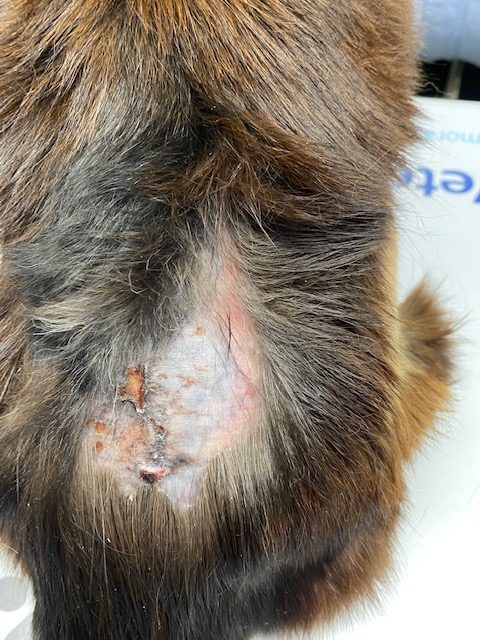
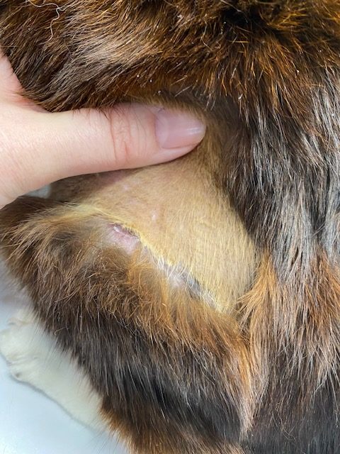
Figures 1 & 2 (above). Thin, multifocally ruptured skin from the Domestic Short-Hair cat in Case 1.
Case 2
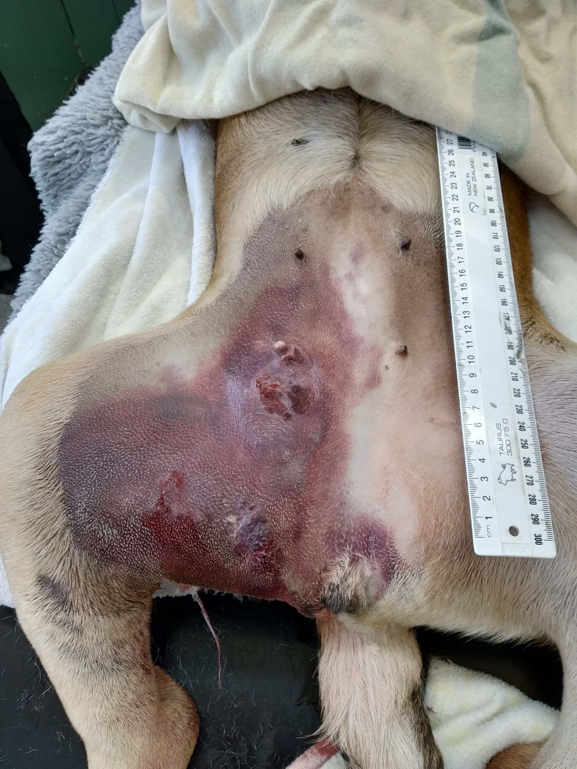
A four-year-old Pig-Dog had a rapidly developing, firm irregular haemorrhagic mass in the right caudal mammary region, with bruised and necrotic-appearing overlying skin and bloody fluid expressible from a nipple.
Initial skin biopsies showed only mild and non-specific reactive and inflammatory changes with oedema. An image was then submitted showing the extent of the lesion (Figure 3) and it was discussed that the initial biopsies were probably not representative of the disease process.

Since the lesion was aggressive and the patient did not respond to supportive treatment, it was euthanased. Subsequent histopathology of larger and deeper samples identified a malignant round cell tumour alongside a concomitant haemangiosarcoma.
How to submit images
To submit images, you may send a hard copy/print out with the submission form, or (preferably) email images to your local laboratory (these email addresses can be found at any time on our website or inside out price book):
> Auckland.vetlab@gribbles.co.nz
> Hamilton.vetlab@gribbles.co.nz
> Palmerston.vetlab@gribbles.co.nz
> Christchurch.vetlab@gribbles.co.nz
> Dunedin.vetlab@gribbles.co.nz
If they are large files or a series of images is being provided (e.g. a CT study), then a link to an external shared drive or online server can alternatively be emailed e.g. Dropbox or similar. We have internal network image storage and document recording systems for linking your email to your submission, allowing any pathologist to review an image when they are examining a case.