
CRISTINA GANS
Clinical history:
A five-year-old, female Rottweiler/ Labrador cross presented for a routine ovariohysterectomy. During the surgery a 5cm mass was identified in the left ovary. The entire uterus and both ovaries were submitted for histopathology.
Post-mortem findings:
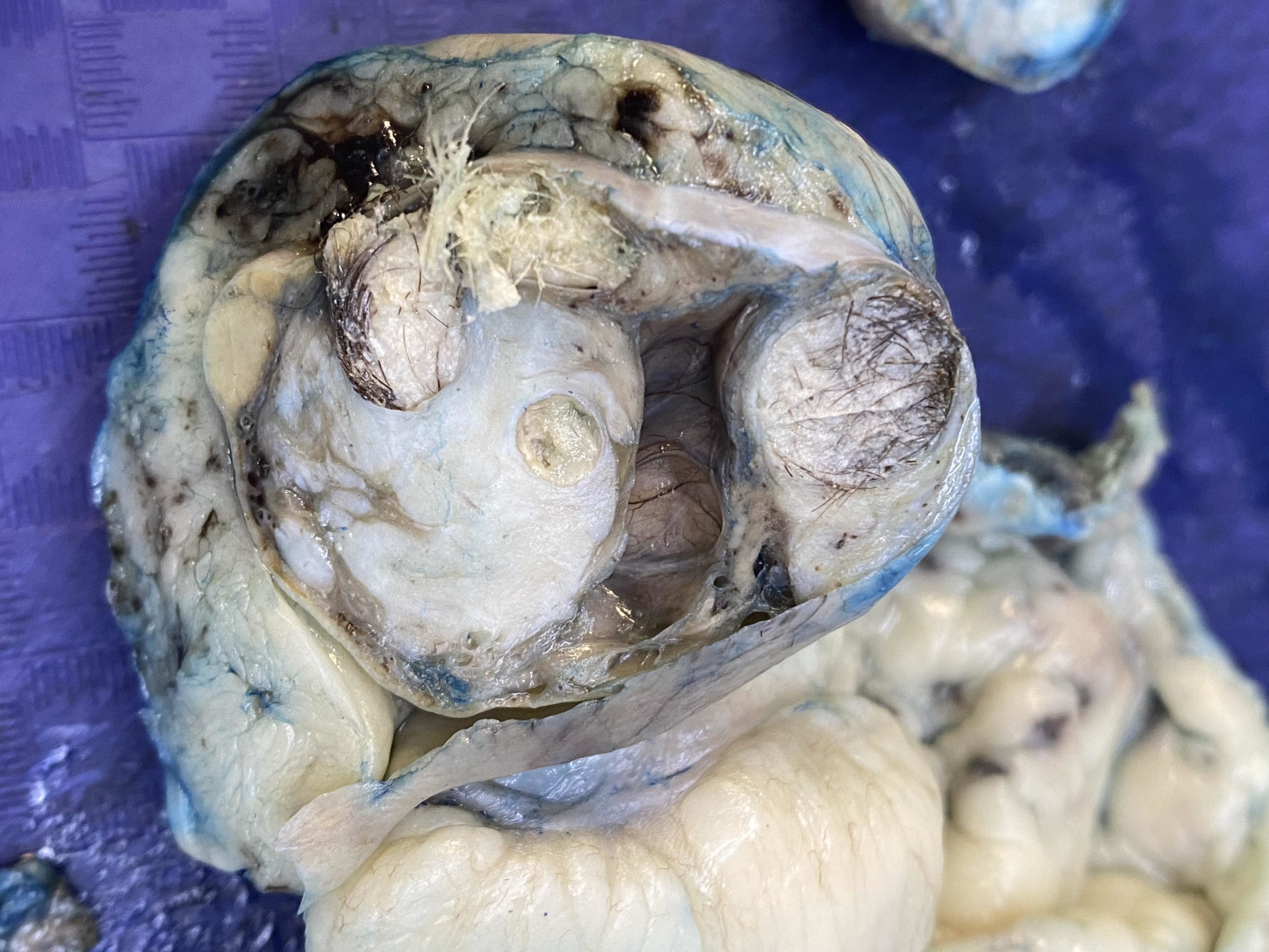
After formalin fixation of the tissue, incision into the ovarian mass, revealed a mixture of tissues including adipose and fibrous tissues and cysts with hair or pale yellow pasty material suggestive of keratin (Figure 1).

Pathology findings:
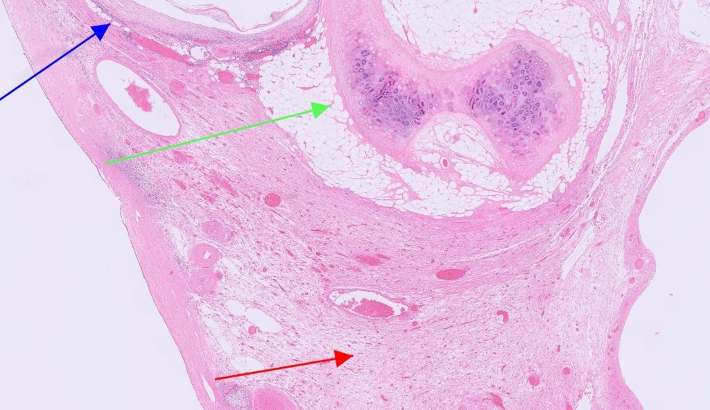
Histological examination of the left ovary revealed a circumscribed neoplasm compressing a narrow rim of normal ovarian tissue. Within the neoplasm were large areas of normal adipose and fibrous connective tissue as well as smaller areas of cartilage, glands lined by ciliated respiratory epithelium and nervous tissue with primitive neurons. Also present were large cystic follicles containing large numbers of hair shafts and keratin (Figure 2).
The remainder of the uterus and right ovary displayed no significant findings.
Diagnosis:
Ovarian teratoma
Discussion:
The presence of a neoplasm with tissues developing from two-three germ layers in a reproductive organ is consistent with a teratoma. The word “teratoma” is derived from the ancient Greek word “teratos” meaning monster.
These tumours arise from totipotential germ cells, which can produce a number of tissues including bone, cartilage, teeth or hair. While they are most commonly reported in the bitch, they comprise less than 1% of canine ovarian tumours, and are thus considered to be very rare. The vast majority of these tumours are benign, although rare malignant variants have been reported.
Thank you to Ashlin Urieli from Selwyn Rakaia Vet Services for this interesting case.